9QUB | pdb_00009qub

Cryo-EM structure of the human NHA2-Fab complex


Experimental Data Snapshot

  • Method: ELECTRON MICROSCOPY
  • Resolution: 2.70 Å
  • Aggregation State: PARTICLE 
  • Reconstruction Method: SINGLE PARTICLE 

wwPDB Validation 3D Report Full Report

Validation slider image for 9QUB

This is version 1.1 of the entry. See complete history

Literature

Structure and Inhibition of the Human Na + /H + Exchanger SLC9B2.

Jung, S.Kokane, S.Li, H.Iwata, S.Nomura, N.Drew, D.

(2025) Int J Mol Sci 26

  • DOI: https://doi.org/10.3390/ijms26094221
  • Primary Citation Related Structures: 
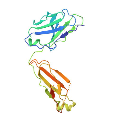
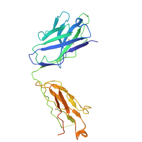
    9QUB, 9QUW

  • PubMed Abstract: 

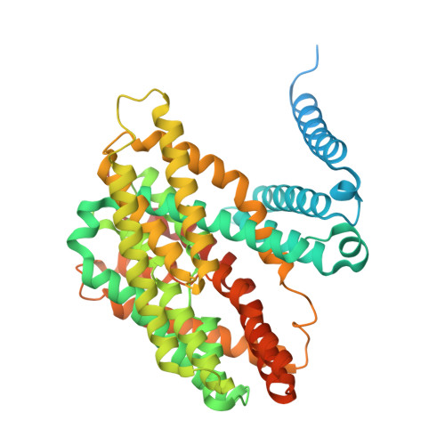
    The sodium/proton exchanger NHA2, also known as SLC9B2, is important for insulin secretion, renal blood pressure regulation, and electrolyte retention. Recent structures of bison NHA2 has revealed its unique 14-transmembrane helix architecture, which is different from SLC9A/NHE members made up from 13-TM helices. Sodium/proton exchangers are functional homodimers, and the additional N-terminal helix in NHA2 was found to alter homodimer assembly. Here, we present the cryo-electron microscopy structures of apo human NHA2 in complex with a Fab fragment and also with the inhibitor phloretin bound at 2.8 and 2.9 Å resolution, respectively. We show how phosphatidic acid (PA) lipids bind to the homodimer interface of NHA2 on the extracellular side, which we propose has a regulatory role linked to cell volume regulation. The ion binding site of human NHA2 has a salt bridge interaction between the ion binding aspartate D278 and R432, an interaction previously broken in the bison NHA2 structure, and these differences suggest a possible ion coupling mechanism. Lastly, the human NHA2 structure in complex with phloretin offers a template for structure-guided drug design, potentially leading to the development of more selective and potent NHA2 inhibitors.


  • Organizational Affiliation
    • Department of Biochemistry and Biophysics, Science for Life Laboratory, Stockholm University, 171-65 Stockholm, Sweden.

Macromolecule Content 

  • Total Structure Weight: 219.47 kDa 
  • Atom Count: 12,879 
  • Modeled Residue Count: 1,685 
  • Deposited Residue Count: 2,014 
  • Unique protein chains: 3

Macromolecules

Find similar proteins by:|  3D Structure
Entity ID: 1
MoleculeChains  Sequence LengthOrganismDetailsImage
Fab-light chainA [auth C],
B [auth E]
214Mus musculusMutation(s): 0 
Find similar proteins by:|  3D Structure
Entity ID: 2
MoleculeChains  Sequence LengthOrganismDetailsImage
Fab-heavy chainC [auth D],
D [auth F]
256Mus musculusMutation(s): 0 
Find similar proteins by:|  3D Structure
Entity ID: 3
MoleculeChains  Sequence LengthOrganismDetailsImage
Sodium/hydrogen exchanger 9B2E [auth B],
F [auth A]
537Homo sapiensMutation(s): 0 
Gene Names: SLC9B2NHA2NHEDC2
Membrane Entity: Yes 
UniProt & NIH Common Fund Data Resources
Find proteins for Q86UD5 (Homo sapiens)
Explore Q86UD5 
Go to UniProtKB:  Q86UD5
PHAROS:  Q86UD5
GTEx:  ENSG00000164038 
Entity Groups
Sequence Clusters30% Identity50% Identity70% Identity90% Identity95% Identity100% Identity
UniProt GroupQ86UD5
Sequence Annotations
Expand
Reference Sequence

Experimental Data & Validation

Experimental Data

  • Method: ELECTRON MICROSCOPY
  • Resolution: 2.70 Å
  • Aggregation State: PARTICLE 
  • Reconstruction Method: SINGLE PARTICLE 
EM Software:
TaskSoftware PackageVersion
MODEL REFINEMENTPHENIX

Structure Validation

View Full Validation Report


Entry History 

& Funding Information

Deposition Data


Funding OrganizationLocationGrant Number
European Research Council (ERC)European Union820187

Revision History  (Full details and data files)

  • Version 1.0: 2025-05-14
    Type: Initial release
  • Version 1.1: 2025-05-28
    Changes: Data collection, Database references