Targeting the Rift Valley Fever Virus Polymerase: Resistance Mechanisms and Structural Insights.
Kral', M., Das, A., Kotacka, T., Blahosova, A., Liscakova, V., Hodek, J., Konvalinka, J., Demo, G., Kozisek, M.(2025) ACS Infect Dis 11: 3364-3376
- PubMed: 41166549 Search on PubMed
- DOI: https://doi.org/10.1021/acsinfecdis.5c00832
- Primary Citation Related Structures:
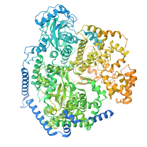
9QTB - PubMed Abstract:
Rift Valley fever virus (RVFV) is an arbovirus from the Phenuiviridae family that can cause severe disease in humans and livestock, with outbreaks resulting in substantial economic losses. Despite the availability of attenuated vaccines for animals, there is no approved preventive or therapeutic agent for human RVFV infections. Moreover, the safety and efficacy of the current veterinary vaccines remain uncertain. The RVFV L protein, a 250 kDa polymerase, plays a key role in viral replication and transcription, containing endonuclease, RNA-dependent RNA polymerase (RdRp), and cap-binding domains. Structurally conserved across related viruses and functionally analogous to the influenza virus polymerase, the L protein is a compelling antiviral target. In our study, we screened a library of polymerase inhibitors and identified several compounds with inhibitory activity against the RVFV polymerase. We validated their effect using both live virus assays and a minigenome luciferase reporter system. Resistance mutants were generated, and key mutations conferring resistance to the inhibitors were identified and characterized. Some of these key mutations were structurally analyzed via cryo-electron microscopy, using a new structure of the apo form of wild-type RVFV L protein resolved at 3.5 Å. This structure provides critical insights into how the mutations can influence inhibitor binding and RVFV polymerase function. These findings provide insight into how these mutations may confer resistance by affecting inhibitor binding and polymerase activity.
- Institute of Organic Chemistry and Biochemistry of the Czech Academy of Sciences, Flemingovo n. 2, 166 10 Prague 6, Czech Republic.
Organizational Affiliation: