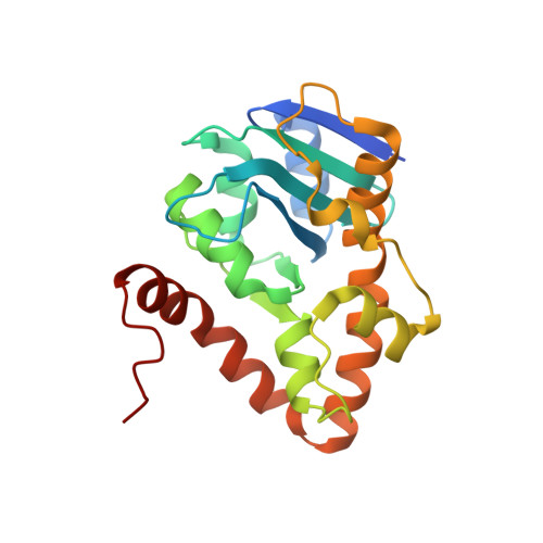
9QRF | pdb_00009qrf

Ancestral protein of diDNase


Experimental Data Snapshot

  • Method: X-RAY DIFFRACTION
  • Resolution: 1.45 Å
  • R-Value Free: 
    0.200 (Depositor), 0.202 (DCC) 
  • R-Value Work: 
    0.166 (Depositor), 0.166 (DCC) 
  • R-Value Observed: 
    0.167 (Depositor) 

Starting Model: in silico
View more details

wwPDB Validation 3D Report Full Report

Validation slider image for 9QRF

This is version 1.1 of the entry. See complete history

Literature

Ancestral proteins trace the emergence of substrate specificity and oligomerization within bacterial DEDDy dinucleases.

Mortensen, S.Burnim, A.A.Dufault-Thompson, K.Lipka, A.E.Jiang, X.Sondermann, H.

(2026) Sci Adv 12: eaef1581-eaef1581

  • DOI: https://doi.org/10.1126/sciadv.aef1581
  • Primary Citation Related Structures: 
    9QRF, 9QTH, 9RTC

  • PubMed Abstract: 

    Nucleases are crucial for various bacterial processes, including genome maintenance and host defense. Deoxydinucleases (diDNases), a class of Gram-positive bacteria-specific nucleases associated with mobile genetic elements, are homologous to nanoRNase C (NrnC) in Gram-negative bacteria but exhibit notable differences: diDNases form dimers and cleave DNA dinucleotides, whereas NrnC forms octamers that process both RNA and DNA dinucleotides. The mechanism by which substrate specificity emerged, and whether it is linked to oligomerization, remained unknown. Here, we reconstructed a common ancestor of diDNases and NrnC orthologs that forms a dimer with intermediate preference for DNA. Structures of ancestral and extant dinucleases reveal gradual changes in conformation that gave rise to substrate preference, oligomeric state, and catalytic efficiency. These findings highlight how subtle, concerted structural modifications enable large-scale changes in molecular assembly and functional specialization, harnessing a conserved protein fold. DNA dinucleotide preference in the early ancestor and preservation of DNase activity in all extant enzymes strongly argue for a biological function of DNA dinucleotides.


  • Organizational Affiliation
    • CSSB Centre for Structural Systems Biology, Deutsches Elektronen-Synchrotron DESY, Notkestr. 85, 22607 Hamburg, Germany.

Macromolecule Content 

  • Total Structure Weight: 24.17 kDa 
  • Atom Count: 1,802 
  • Modeled Residue Count: 202 
  • Deposited Residue Count: 211 
  • Unique protein chains: 1

Macromolecules

Find similar proteins by:|  3D Structure
Entity ID: 1
MoleculeChains  Sequence LengthOrganismDetailsImage
ancestor of diDNase211synthetic constructMutation(s): 0 
Entity Groups
Sequence Clusters30% Identity50% Identity70% Identity90% Identity95% Identity100% Identity
Sequence Annotations
Expand
Reference Sequence

Small Molecules

Ligands 1 Unique
IDChains Name / Formula / InChI Key2D Diagram3D Interactions
CL

Query on CL


Download:Ideal Coordinates CCD File
B [auth A]CHLORIDE ION
Cl
VEXZGXHMUGYJMC-UHFFFAOYSA-M

Experimental Data & Validation

Experimental Data

  • Method: X-RAY DIFFRACTION
  • Resolution: 1.45 Å
  • R-Value Free:  0.200 (Depositor), 0.202 (DCC) 
  • R-Value Work:  0.166 (Depositor), 0.166 (DCC) 
  • R-Value Observed: 0.167 (Depositor) 
Space Group: C 1 2 1
Unit Cell:
Length ( Å )Angle ( ˚ )
a = 70.903α = 90
b = 70.948β = 115.807
c = 48.241γ = 90
Software Package:
Software NamePurpose
PHENIXrefinement
PHENIXrefinement
XDSdata reduction
XDSdata scaling
PHASERphasing

Structure Validation

View Full Validation Report


Entry History 

& Funding Information

Deposition Data


Funding OrganizationLocationGrant Number
National Institutes of Health/National Human Genome Research Institute (NIH/NHGRI)United StatesR01 AI142400

Revision History  (Full details and data files)

  • Version 1.0: 2026-04-15
    Type: Initial release
  • Version 1.1: 2026-08-26
    Changes: Database references