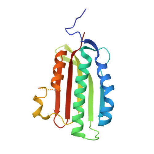
9QL9 | pdb_00009ql9

CA domain of LvrB from Leptospira in its apo form


Experimental Data Snapshot

  • Method: X-RAY DIFFRACTION
  • Resolution: 1.50 Å
  • R-Value Free: 
    0.237 (Depositor), 0.237 (DCC) 
  • R-Value Work: 
    0.195 (Depositor), 0.196 (DCC) 
  • R-Value Observed: 
    0.198 (Depositor) 

Starting Model: experimental
View more details

wwPDB Validation 3D Report Full Report

Validation slider image for 9QL9

This is version 1.1 of the entry. See complete history. 

Literature

Activation mechanism of the full-length histidine kinase LvrB from pathogenic Leptospira.

Agustoni, E., Mechaly, A., Dalla Rizza, J., Beriashvili, D., Pluhackova, K., Isaikina, P., Trajtenberg, F., Muntener, T., Wunder Jr., E.A., Ko, A.I., Schirmer, T., Buschiazzo, A., Hiller, S.

(2026) Nat Commun 17

  • DOI: https://doi.org/10.1038/s41467-026-71783-4
  • Primary Citation Related Structures: 
    8VC9, 9QIH, 9QJG, 9QL9, 9QQW, 9QR2

  • PubMed Abstract: 

    Pathogenic Leptospira modulate their virulence via the Lvr signaling system, with the histidine kinase LvrB being a central element. LvrB is a prototype of Rec-controlled histidine kinases, which are frequently found in bacterial two-component systems, and yet whose regulatory mechanisms remain largely unknown. Here, we report full-length structures of LvrB in different states uncovering its mechanism of activation. Kinase-inactive LvrB is a symmetric homodimer, with its catalytic domains rigidly clasped onto the central helical domain. Phosphorylation of the N-terminal Rec domains induces coiled-coil formation of the central αS helices thereby breaking symmetry through liberation of the catalytic domains into a dynamic, auto-phosphorylation competent state. We further identified LvrB's downstream effector partner LvrC, an anti-σ factor that reprograms the transcription of hundreds of virulence genes. Our findings set a mechanistic paradigm for Rec-controlled histidine kinases enabling the design of virulence inhibitors.


  • Organizational Affiliation: 
    • Biozentrum, University of Basel, Basel, Switzerland.

Macromolecule Content 

  • Total Structure Weight: 39.33 kDa 
  • Atom Count: 2,639 
  • Modeled Residue Count: 302 
  • Deposited Residue Count: 346 
  • Unique protein chains: 1

Macromolecules

Find similar proteins by:|  3D Structure
Entity ID: 1
MoleculeChains  Sequence LengthOrganismDetailsImage
histidine kinase
A, B
173Leptospira interrogansMutation(s): 0 
Gene Names: LA_2223
EC: 2.7.13.3
UniProt
Find proteins for Q8F424 (Leptospira interrogans serogroup Icterohaemorrhagiae serovar Lai (strain 56601))
Explore Q8F424 
Go to UniProtKB:  Q8F424
Entity Groups
Sequence Clusters30% Identity50% Identity70% Identity90% Identity95% Identity100% Identity
UniProt GroupQ8F424
Sequence Annotations
Expand
Reference Sequence

Experimental Data & Validation

Experimental Data

  • Method: X-RAY DIFFRACTION
  • Resolution: 1.50 Å
  • R-Value Free:  0.237 (Depositor), 0.237 (DCC) 
  • R-Value Work:  0.195 (Depositor), 0.196 (DCC) 
  • R-Value Observed: 0.198 (Depositor) 
Space Group: P 21 21 21
Unit Cell:
Length ( Å )Angle ( ˚ )
a = 41.543α = 90
b = 57.153β = 90
c = 135.448γ = 90
Software Package:
Software NamePurpose
PHENIXrefinement
XDSdata reduction
Aimlessdata scaling
PHASERphasing

Structure Validation

View Full Validation Report


Entry History 

& Funding Information

Deposition Data


Funding OrganizationLocationGrant Number
Novartis FreeNovationSwitzerland--

Revision History  (Full details and data files)

  • Version 1.0: 2026-04-08
    Type: Initial release
  • Version 1.1: 2026-10-07
    Changes: Database references