


Structure of E. coli IspH crystallized in the presence of adenosine hemisulfate
Bikbaev, K., Dormann, C., Span, I.To be published.
Experimental Data Snapshot
Starting Model: experimental
View more details
Entity ID: 1 | |||||
|---|---|---|---|---|---|
| Molecule | Chains | Sequence Length | Organism | Details | Image |
| 4-hydroxy-3-methylbut-2-enyl diphosphate reductase | 327 | Escherichia coli K-12 | Mutation(s): 0 Gene Names: ispH, lytB, yaaE, b0029, JW0027 EC: 1.17.7.4 | 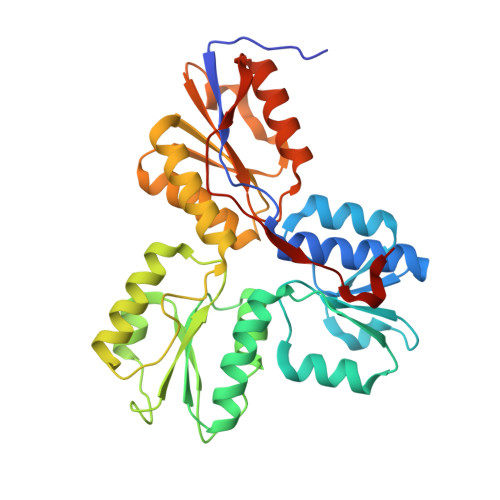 | |
UniProt | |||||
Entity Groups | |||||
| Sequence Clusters | 30% Identity50% Identity70% Identity90% Identity95% Identity100% Identity | ||||
| UniProt Group | P62623 | ||||
Sequence AnnotationsExpand | |||||
Reference Sequence | |||||
| Ligands 2 Unique | |||||
|---|---|---|---|---|---|
| ID | Chains | Name / Formula / InChI Key | 2D Diagram | 3D Interactions | |
| SF4 (Subject of Investigation/LOI) Download:Ideal Coordinates CCD File | C [auth A], E [auth B] | IRON/SULFUR CLUSTER Fe4 S4 LJBDFODJNLIPKO-UHFFFAOYSA-N |  | ||
| SO4 (Subject of Investigation/LOI) Download:Ideal Coordinates CCD File | D [auth A], F [auth B] | SULFATE ION O4 S QAOWNCQODCNURD-UHFFFAOYSA-L |  | ||
| Length ( Å ) | Angle ( ˚ ) |
|---|---|
| a = 67.909 | α = 90 |
| b = 81.291 | β = 90 |
| c = 112.871 | γ = 90 |
| Software Name | Purpose |
|---|---|
| REFMAC | refinement |
| Coot | model building |
| XDS | data reduction |
| Aimless | data scaling |
| MOLREP | phasing |
| Funding Organization | Location | Grant Number |
|---|---|---|
| German Research Foundation (DFG) | Germany | SP 1476/4-1 |


