9QKY | pdb_00009qky

The structure of the DNA-binding domain of Nuclear Factor 1 X bound to NFI consensus DNA sequence


Experimental Data Snapshot

  • Method: ELECTRON MICROSCOPY
  • Resolution: 3.86 Å
  • Aggregation State: FILAMENT 
  • Reconstruction Method: HELICAL 

Starting Model: experimental
View more details

wwPDB Validation 3D Report Full Report

Validation slider image for 9QKY

This is version 1.1 of the entry. See complete history

Literature

Structural basis of Nuclear Factor 1-X DNA recognition provides prototypic insight into the NFI family.

Tiberi, M.Lapi, M.Gourlay, L.J.Chaves-Sanjuan, A.Polentarutti, M.Demitri, N.Cavinato, M.Bonnet, D.M.V.Taglietti, V.Righetti, A.Sala, R.Cauteruccio, S.Kumawat, A.Russo, R.Barbiroli, A.G.Gnesutta, N.Camilloni, C.Bolognesi, M.Messina, G.Nardini, M.

(2025) Nat Commun 16: 10170-10170

  • DOI: https://doi.org/10.1038/s41467-025-65186-0
  • Primary Citation Related Structures: 
    7QQD, 9QKY

  • PubMed Abstract: 

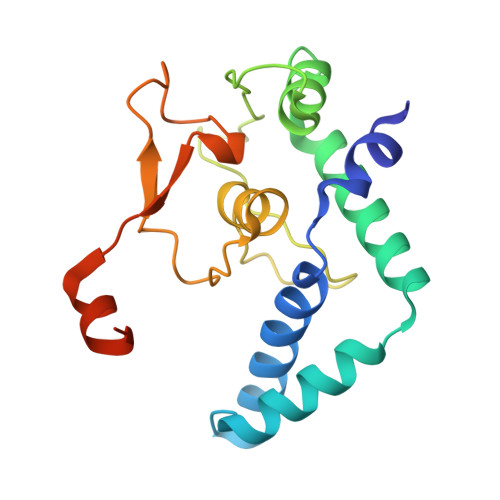
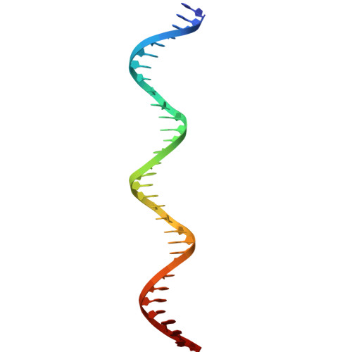
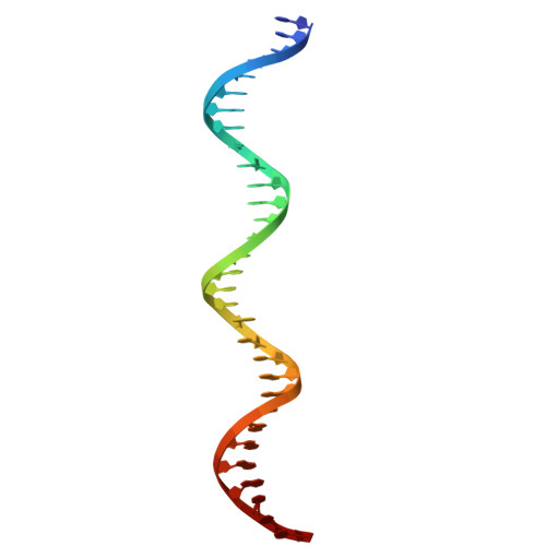
    Nuclear Factor I (NFI) proteins are involved in adenovirus DNA replication and regulate gene transcription, stem cell proliferation, and differentiation. They play key roles in development, cancer, and congenital disorders. Within the NFI family, NFI-X is critical for neural stem cell biology, hematopoiesis, muscle development, muscular dystrophies, and oncogenesis. Here, we present the structural characterization of the NFI transcription factor NFI-X, both alone and bound to its consensus palindromic DNA site. Our analyses reveal a MH1-like fold within NFI-X DNA-binding domain (DBD) and identify crucial structural determinants for activity, such as a Zn²⁺ binding site, dimeric assembly, and DNA-binding specificity. Given the ~85% sequence identity within the NFI DBDs, our structural data are prototypic for the entire family, a NFI Rosetta Stone that allows decoding a wealth of biochemical and functional data and provides a precise target for drug design in a wider disease context.


  • Organizational Affiliation
    • Department of Biosciences, Università degli Studi di Milano, Milano, Italy.

Macromolecule Content 

  • Total Structure Weight: 247.43 kDa 
  • Atom Count: 16,276 
  • Modeled Residue Count: 1,600 
  • Deposited Residue Count: 1,704 
  • Unique protein chains: 1
  • Unique nucleic acid chains: 2

Macromolecules


Find similar proteins by:|  3D Structure
Entity ID: 1
MoleculeChains  Sequence LengthOrganismDetailsImage
Nuclear factor 1 X-type
A, B, E, F, I
A, B, E, F, I, J, M, N
182Mus musculusMutation(s): 0 
Gene Names: Nfix
UniProt
Find proteins for P70257 (Mus musculus)
Explore P70257 
Go to UniProtKB:  P70257
Entity Groups
Sequence Clusters30% Identity50% Identity70% Identity90% Identity95% Identity100% Identity
UniProt GroupP70257
Sequence Annotations
Expand
Reference Sequence
Find similar nucleic acids by:  (by identity cutoff) 
Entity ID: 2
MoleculeChains LengthOrganismImage
DNA (31-MER)
C, G, K, O
31Mus musculus
Sequence Annotations
Expand
Reference Sequence
Find similar nucleic acids by:  (by identity cutoff) 
Entity ID: 3
MoleculeChains LengthOrganismImage
DNA (31-MER)
D, H, L, P
31Mus musculus
Sequence Annotations
Expand
Reference Sequence

Experimental Data & Validation

Experimental Data

  • Method: ELECTRON MICROSCOPY
  • Resolution: 3.86 Å
  • Aggregation State: FILAMENT 
  • Reconstruction Method: HELICAL 
EM Software:
TaskSoftware PackageVersion
RECONSTRUCTIONcryoSPARC
MODEL REFINEMENTPHENIX1.20.1_4487

Structure Validation

View Full Validation Report


Entry History 

& Funding Information

Deposition Data


Funding OrganizationLocationGrant Number
Not funded--

Revision History  (Full details and data files)

  • Version 1.0: 2025-09-17
    Type: Initial release
  • Version 1.1: 2025-12-24
    Changes: Data collection, Database references