9PUZ | pdb_00009puz

HRAS complex with UM0153030 compound


Experimental Data Snapshot

  • Method: X-RAY DIFFRACTION
  • Resolution: 2.00 Å
  • R-Value Free: 
    0.253 (Depositor), 0.255 (DCC) 
  • R-Value Work: 
    0.209 (Depositor), 0.210 (DCC) 
  • R-Value Observed: 
    0.212 (Depositor) 

Starting Model: experimental
View more details

wwPDB Validation 3D Report Full Report

Validation slider image for 9PUZ

Ligand Structure Quality Assessment 


This is version 1.1 of the entry. See complete history

Literature

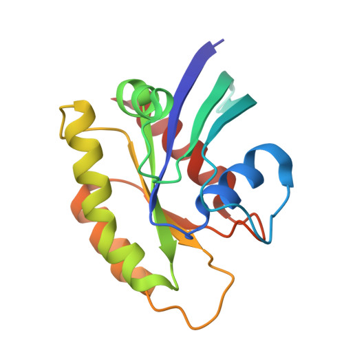
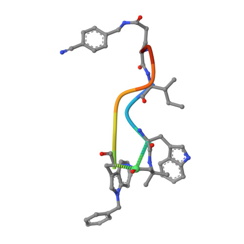
Targeting the H/KRAS alpha 4-beta 6-alpha 5 Allosteric Lobe with Macrocyclic Peptides.

Tran, K.Lavoie, H.Wahhab, A.Garrido, D.Jo, C.H.Poupart, M.A.Arya, T.Beautrait, A.Killoran, R.Dicaire-Leduc, C.Bonneil, E.Osborne, M.Schuetz, D.A.Shaikh, F.Thibault, P.Smith, M.J.Marinier, A.Therrien, M.

(2026) ACS Med Chem Lett 17: 1154-1162

  • DOI: https://doi.org/10.1021/acsmedchemlett.6c00078
  • Primary Citation Related Structures: 
    9PU1, 9PU3, 9PU8, 9PUL, 9PUN, 9PUQ, 9PUT, 9PUZ, 9PVE, 9PVF

  • PubMed Abstract: 

    Despite therapeutic advances against RAS mutations in cancer, acquired resistance frequently arises. Several secondary mutations at the binding sites effectively confer resistance to both Switch-II inhibitors and cyclophilin-A molecular glues. This underscores the need for RAS inhibitors that engage alternative binding pockets or operate through novel mechanisms. Here, we report the design of 10-mer macrocyclic peptides that mimic the FG-loop of the NS1 monobody, which targets the allosteric α4-α5-β6 surface of H/KRAS to disrupt RAS clustering and downstream signaling. These noncovalent inhibitors bind to H/KRAS with equivalent potencies, regardless of nucleotide state or the presence of oncogenic mutations (G12D, G12V, G13R, Q61K), and their binding site was confirmed by NMR and X-ray crystallography. Furthermore, covalent analogs targeting Cys118 were shown to label RAS in vitro and in complete cell lysates. Finally, we demonstrated that the key pharmacophores are connectable, providing a foundation for the development of smaller allosteric H/KRAS inhibitors.


  • Organizational Affiliation
    • Institute for Research in Immunology and Cancer, Université de Montréal, Montréal, QC H3C 3J7, Canada.

Macromolecule Content 

  • Total Structure Weight: 63.33 kDa 
  • Atom Count: 4,594 
  • Modeled Residue Count: 531 
  • Deposited Residue Count: 531 
  • Unique protein chains: 2

Macromolecules

Find similar proteins by:|  3D Structure
Entity ID: 1
MoleculeChains  Sequence LengthOrganismDetailsImage
GTPase HRas
A, B, C
166Homo sapiensMutation(s): 0 
Gene Names: HRASHRAS1
EC: 3.6.5.2
UniProt & NIH Common Fund Data Resources
Find proteins for P01112 (Homo sapiens)
Explore P01112 
Go to UniProtKB:  P01112
PHAROS:  P01112
GTEx:  ENSG00000174775 
Entity Groups
Sequence Clusters30% Identity50% Identity70% Identity90% Identity95% Identity100% Identity
UniProt GroupP01112
Sequence Annotations
Expand
Reference Sequence
Find similar proteins by:  Sequence   |   3D Structure  
Entity ID: 2
MoleculeChains  Sequence LengthOrganismDetailsImage
UM0153030D [auth E],
E [auth F],
F [auth G]
11synthetic constructMutation(s): 0 

Small Molecules

Ligands 3 Unique
IDChains Name / Formula / InChI Key2D Diagram3D Interactions
GDP

Query on GDP


Download:Ideal Coordinates CCD File
H [auth A],
L [auth B],
O [auth C]
GUANOSINE-5'-DIPHOSPHATE
C10 H15 N5 O11 P2
QGWNDRXFNXRZMB-UUOKFMHZSA-N
PO4

Query on PO4


Download:Ideal Coordinates CCD File
I [auth A],
J [auth A],
M [auth B],
P [auth C]
PHOSPHATE ION
O4 P
NBIIXXVUZAFLBC-UHFFFAOYSA-K
MG

Query on MG


Download:Ideal Coordinates CCD File
G [auth A],
K [auth B],
N [auth C]
MAGNESIUM ION
Mg
JLVVSXFLKOJNIY-UHFFFAOYSA-N
Modified Residues  2 Unique
IDChains TypeFormula2D DiagramParent
A1CK7
Query on A1CK7
D [auth E],
E [auth F],
F [auth G]
PEPTIDE-LIKEC5 H11 N O2

--

A1CLH
Query on A1CLH
D [auth E],
E [auth F],
F [auth G]
D-PEPTIDE LINKINGC18 H18 N2 O2TRP
DPR
Query on DPR
D [auth E],
E [auth F],
F [auth G]
D-PEPTIDE LINKINGC5 H9 N O2

--

IIL
Query on IIL
D [auth E],
E [auth F],
F [auth G]
L-PEPTIDE LINKINGC6 H13 N O2ILE

Experimental Data & Validation

Experimental Data

  • Method: X-RAY DIFFRACTION
  • Resolution: 2.00 Å
  • R-Value Free:  0.253 (Depositor), 0.255 (DCC) 
  • R-Value Work:  0.209 (Depositor), 0.210 (DCC) 
  • R-Value Observed: 0.212 (Depositor) 
Space Group: P 63
Unit Cell:
Length ( Å )Angle ( ˚ )
a = 85.72α = 90
b = 85.72β = 90
c = 136.27γ = 120
Software Package:
Software NamePurpose
PHENIXrefinement
Cootmodel building

Structure Validation

View Full Validation Report


Ligand Structure Quality Assessment 


Entry History 

& Funding Information

Deposition Data


Funding OrganizationLocationGrant Number
Not funded--

Revision History  (Full details and data files)

  • Version 1.0: 2026-04-22
    Type: Initial release
  • Version 1.1: 2026-06-03
    Changes: Database references