9PUO | pdb_00009puo

Neutralizing monoclonal antibody Fab fragment bound to leptin

  • Classification: IMMUNE SYSTEM
  • Organism(s): Homo sapiens
  • Expression System: Homo sapiens, Escherichia coli BL21(DE3)
  • Mutation(s): Yes 

  • Deposited: 2025-07-31 Released: 2025-11-05 
  • Deposition Author(s): Tomchick, D.R., Wynn, R.M., Scherer, P.E.
  • Funding Organization(s): National Institutes of Health/National Institute of Diabetes and Digestive and Kidney Disease (NIH/NIDDK), National Institutes of Health/National Institute on Aging (NIH/NIA), Cancer Prevention and Research Institute of Texas (CPRIT), Robert A. Welch Foundation

Experimental Data Snapshot

  • Method: X-RAY DIFFRACTION
  • Resolution: 3.10 Å
  • R-Value Free: 
    0.277 (Depositor), 0.277 (DCC) 
  • R-Value Work: 
    0.236 (Depositor), 0.236 (DCC) 
  • R-Value Observed: 
    0.240 (Depositor) 

Starting Models: experimental
View more details

wwPDB Validation 3D Report Full Report

Validation slider image for 9PUO

This is version 1.0 of the entry. See complete history

Literature

Leptin as a key driver for organ fibrogenesis.

Sun, X.N.Chen, S.Zhao, S.Funcke, J.B.Virostek, M.Pedersen, L.Li, C.Joung, C.Lin, Q.Li, Y.Cobb, A.Wang, M.Y.Min, K.Maya-Ramos, L.Degasperi, G.Liu, J.Zhang, N.An, Z.Tomchick, D.R.Wynn, R.M.Oh, D.Y.Scherer, P.E.

(2025) Sci Adv 11: eady7904-eady7904

  • DOI: https://doi.org/10.1126/sciadv.ady7904
  • Primary Citation Related Structures: 
    9PUK, 9PUO

  • PubMed Abstract: 

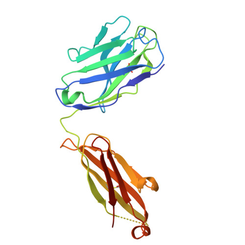
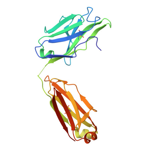
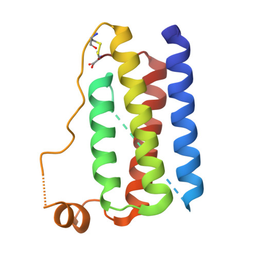
    Leptin, a hormone primarily secreted by adipocytes, regulates energy balance and systemic metabolism through its interaction with the leptin receptor (LEPR). Beyond these functions, leptin signaling has been implicated in the pathogenesis of tissue fibrosis. Here, we report the x-ray crystal structures of a leptin-neutralizing antibody (hLep3) in the unbound and leptin-bound states. The interaction of this antibody with leptin mimics the interaction of the LEPR with leptin, providing direct insights into the mechanism by which the antibody disrupts leptin signaling. We furthermore evaluate the therapeutic potential of neutralizing leptin with this antibody across distinct mouse models of fibrosis affecting the kidney, liver, lung, heart, and blood vessels. Leptin neutralization markedly inhibited fibrosis progression in all models. Mechanistically, suppression of leptin activity reduces pro-inflammatory and profibrotic processes, underscoring its therapeutic potential. These findings suggest that leptin signaling plays a vital role in tissue fibrosis and that treatment with a leptin-neutralizing antibody may be a promising therapeutic approach.


  • Organizational Affiliation
    • Touchstone Diabetes Center, University of Texas Southwestern Medical Center, Dallas, TX 75390, USA.

Macromolecule Content 

  • Total Structure Weight: 125.39 kDa 
  • Atom Count: 8,152 
  • Modeled Residue Count: 1,084 
  • Deposited Residue Count: 1,176 
  • Unique protein chains: 3

Macromolecules

Find similar proteins by:|  3D Structure
Entity ID: 1
MoleculeChains  Sequence LengthOrganismDetailsImage
Neutralizing antibody Fab fragment, heavy chain
A, D
225Homo sapiensMutation(s): 0 
Find similar proteins by:|  3D Structure
Entity ID: 2
MoleculeChains  Sequence LengthOrganismDetailsImage
Neutralizing antibody Fab fragment, light chain
B, E
217Homo sapiensMutation(s): 0 
Find similar proteins by:|  3D Structure
Entity ID: 3
MoleculeChains  Sequence LengthOrganismDetailsImage
Leptin
C, F
146Homo sapiensMutation(s): 1 
Gene Names: LEPOBOBS
UniProt & NIH Common Fund Data Resources
Find proteins for P41159 (Homo sapiens)
Explore P41159 
Go to UniProtKB:  P41159
PHAROS:  P41159
GTEx:  ENSG00000174697 
Entity Groups
Sequence Clusters30% Identity50% Identity70% Identity90% Identity95% Identity100% Identity
UniProt GroupP41159
Sequence Annotations
Expand
Reference Sequence

Experimental Data & Validation

Experimental Data

  • Method: X-RAY DIFFRACTION
  • Resolution: 3.10 Å
  • R-Value Free:  0.277 (Depositor), 0.277 (DCC) 
  • R-Value Work:  0.236 (Depositor), 0.236 (DCC) 
  • R-Value Observed: 0.240 (Depositor) 
Space Group: P 1
Unit Cell:
Length ( Å )Angle ( ˚ )
a = 51.927α = 90.344
b = 48.534β = 85.206
c = 124.963γ = 70.418
Software Package:
Software NamePurpose
PHENIXrefinement
HKL-3000data scaling
PHASERphasing
PDB_EXTRACTdata extraction
HKL-3000data reduction

Structure Validation

View Full Validation Report


Entry History 

& Funding Information

Deposition Data


Funding OrganizationLocationGrant Number
National Institutes of Health/National Institute of Diabetes and Digestive and Kidney Disease (NIH/NIDDK)United StatesDK55758
National Institutes of Health/National Institute of Diabetes and Digestive and Kidney Disease (NIH/NIDDK)United StatesDK099110
National Institutes of Health/National Institute of Diabetes and Digestive and Kidney Disease (NIH/NIDDK)United StatesDK118620
National Institutes of Health/National Institute of Diabetes and Digestive and Kidney Disease (NIH/NIDDK)United StatesDK127274
National Institutes of Health/National Institute of Diabetes and Digestive and Kidney Disease (NIH/NIDDK)United StatesDK131537
National Institutes of Health/National Institute on Aging (NIH/NIA)United StatesAG051459
National Institutes of Health/National Institute of Diabetes and Digestive and Kidney Disease (NIH/NIDDK)United StatesDK138935
National Institutes of Health/National Institute on Aging (NIH/NIA)United StatesAG084646
Cancer Prevention and Research Institute of Texas (CPRIT)United StatesRP220032
Cancer Prevention and Research Institute of Texas (CPRIT)United StatesRP150551
Cancer Prevention and Research Institute of Texas (CPRIT)United StatesRP190561
Robert A. Welch FoundationUnited StatesAU-0042-20030616

Revision History  (Full details and data files)

  • Version 1.0: 2025-11-05
    Type: Initial release