9PKV | pdb_00009pkv

HU-38 Fab with PRAME pMHC


Experimental Data Snapshot

  • Method: ELECTRON MICROSCOPY
  • Resolution: 2.72 Å
  • Aggregation State: PARTICLE 
  • Reconstruction Method: SINGLE PARTICLE 

wwPDB Validation 3D Report Full Report

Validation slider image for 9PKV

This is version 1.1 of the entry. See complete history

Literature

Development of a PRAME pMHC targeted T cell engager for solid tumor therapy.

Skrzypczynska, K.Schimert, K.Stephenson, H.Mah, I.K.Mortenson, D.Boyd, K.Hardman, T.Novikov, N.Seto, E.Lu, S.Yen, R.Lee, B.Wang, M.Kang, D.Huang, Y.Yu, X.Hung, M.Ding, S.Thomsen, N.Oakdale, N.S.

(2025) MAbs 17: 2563773-2563773

  • DOI: https://doi.org/10.1080/19420862.2025.2563773
  • Primary Citation Related Structures: 
    9PKV

  • PubMed Abstract: 

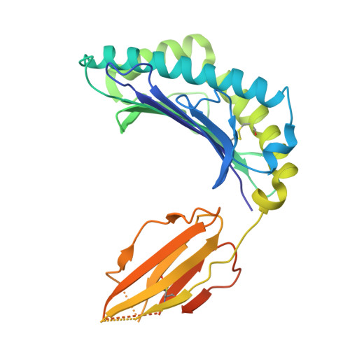
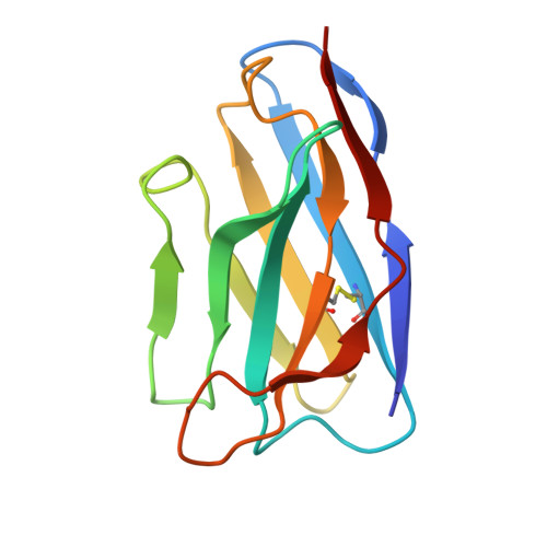
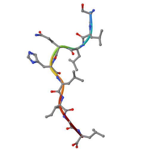
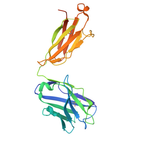
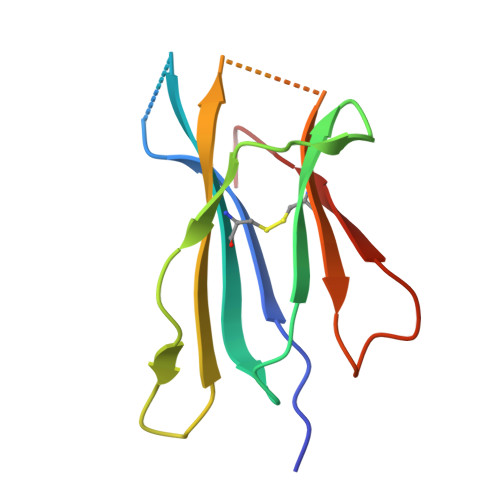
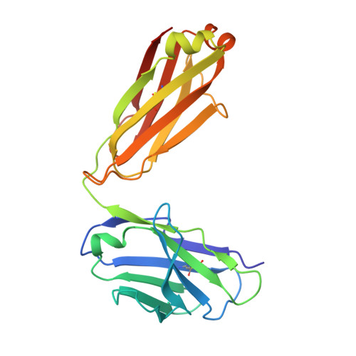
    Bispecific T cell engager (TCE) therapies have demonstrated transformative clinical success in the treatment of hematological cancers, but the lack of antigens that are sufficiently selective for malignant cells has hampered the success of TCEs in the solid-tumor space. To overcome the on-target, off-tumor toxicities that result from the expression of even low levels of tumor-associated antigens in healthy tissues, we sought to identify a TCE target with highly tumor-restricted expression patterns. Here, we characterize cancer-testes antigen Preferentially Expressed Antigen in Melanoma (PRAME) as a highly selective tumor antigen and identify a proteasomal degradation peptide PRAME 425-433 (PRAME 425 ) presented in the context of major histocompatibility complex I (MHCI) as an attractive TCE target. We designed a TCR-mimic (TCRm) antibody screening cascade that prioritizes screening anti-PRAME pMHC binders in off-target T cell dependent cellular cytotoxicity assays in a potent TCE format, rather than relying solely on traditional pMHC binding assays, to determine specificity. Using this screening cascade, we discovered antibodies that selectively bind PRAME 425 pMHC without over-recognition of off-target peptides or MHCI via a TCR-like binding geometry. We further solved the first structure of an anti-PRAME 425 pMHC TCRm antibody in complex with PRAME 425 /HLA-A *02:01 using cryo electron microscopy to confirm the TCRm antibody binds in a TCR-like binding geometry and specifically recognizes the PRAME 425 peptide. By formatting these novel TCRm antibodies into potent TCEs, we demonstrate PRAME 425 pMHC-specific killing of tumor cells, representing a new class of anti-PRAME pMHC biologics.


  • Organizational Affiliation
    • Oncology, Gilead Sciences, Inc, Foster City, CA, USA.

Macromolecule Content 

  • Total Structure Weight: 109.45 kDa 
  • Atom Count: 7,062 
  • Modeled Residue Count: 887 
  • Deposited Residue Count: 982 
  • Unique protein chains: 6

Macromolecules

Find similar proteins by:|  3D Structure
Entity ID: 1
MoleculeChains  Sequence LengthOrganismDetailsImage
MHC class I antigen301Homo sapiensMutation(s): 0 
Gene Names: HLA-A
UniProt
Find proteins for Q8WLS4 (Homo sapiens)
Explore Q8WLS4 
Go to UniProtKB:  Q8WLS4
Entity Groups
Sequence Clusters30% Identity50% Identity70% Identity90% Identity95% Identity100% Identity
UniProt GroupQ8WLS4
Sequence Annotations
Expand
Reference Sequence
Find similar proteins by:|  3D Structure
Entity ID: 2
MoleculeChains  Sequence LengthOrganismDetailsImage
VHH antibodyB [auth K]121Lama glamaMutation(s): 0 
Find similar proteins by:  Sequence   |   3D Structure  
Entity ID: 3
MoleculeChains  Sequence LengthOrganismDetailsImage
PRAME peptideC [auth P]9Homo sapiensMutation(s): 0 
Find similar proteins by:|  3D Structure
Entity ID: 4
MoleculeChains  Sequence LengthOrganismDetailsImage
HU-38 Heavy ChainD [auth H]237Mus musculusMutation(s): 0 
Find similar proteins by:|  3D Structure
Entity ID: 5
MoleculeChains  Sequence LengthOrganismDetailsImage
Beta-2-microglobulinE [auth B]100Homo sapiensMutation(s): 0 
Gene Names: B2MCDABP0092HDCMA22P
UniProt & NIH Common Fund Data Resources
Find proteins for P61769 (Homo sapiens)
Explore P61769 
Go to UniProtKB:  P61769
PHAROS:  P61769
GTEx:  ENSG00000166710 
Entity Groups
Sequence Clusters30% Identity50% Identity70% Identity90% Identity95% Identity100% Identity
UniProt GroupP61769
Sequence Annotations
Expand
Reference Sequence
Find similar proteins by:|  3D Structure
Entity ID: 6
MoleculeChains  Sequence LengthOrganismDetailsImage
HU-38 Light ChainF [auth L]214Mus musculusMutation(s): 0 

Experimental Data & Validation

Experimental Data

  • Method: ELECTRON MICROSCOPY
  • Resolution: 2.72 Å
  • Aggregation State: PARTICLE 
  • Reconstruction Method: SINGLE PARTICLE 
EM Software:
TaskSoftware PackageVersion
MODEL REFINEMENTPHENIX1.21_5207
RECONSTRUCTIONcryoSPARC

Structure Validation

View Full Validation Report


Entry History 

& Funding Information

Deposition Data


Funding OrganizationLocationGrant Number
Not funded--

Revision History  (Full details and data files)

  • Version 1.0: 2025-10-01
    Type: Initial release
  • Version 1.1: 2026-04-29
    Changes: Data collection, Database references