9PI2 | pdb_00009pi2

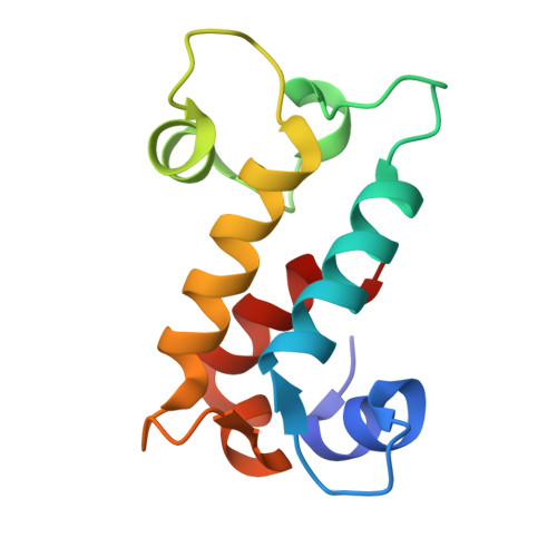
X-ray crystal structure of Ancylobacter lacus LanM bound to Nd(III) and Ca(II)


Experimental Data Snapshot

  • Method: X-RAY DIFFRACTION
  • Resolution: 0.99 Å
  • R-Value Free: 
    0.144 (Depositor), 0.138 (DCC) 
  • R-Value Work: 
    0.136 (Depositor), 0.130 (DCC) 
  • R-Value Observed: 
    0.136 (Depositor) 

wwPDB Validation 3D Report Full Report

Validation slider image for 9PI2

Ligand Structure Quality Assessment 


This is version 1.2 of the entry. See complete history. 

Literature

Near-Adjacent Heavy Lanthanide Separation and Sensing Using Dimerizing Lanmodulins.

Choi, W., Zhou, X., Dong, Z., Song, F., Jung, J.J., Mattocks, J.A., Chlebek, J.L., Diep, P., Johnson, A.C., Crawford, S.E., Jiao, Y., Honaker, R., Boal, A.K., Park, D.M., Cotruvo Jr., J.A.

(2026) J Am Chem Soc 148: 36817-36831

  • DOI: https://doi.org/10.1021/jacs.6c08525
  • Primary Citation Related Structures: 
    9PI2

  • PubMed Abstract: 

    Protein-based recovery, detection, and separation of rare earth elements (REs) have accelerated since the discovery of the lanmodulin (LanM) proteins. While exceptional at total RE recovery, prior proteins, like most ligands, exhibited limited ability to differentiate REs, whether in detection or separations. Here we report two dimerizing LanMs (Al-LanM and Xan-LanM) that address these challenges. Uniquely among biomolecules, these proteins promote luminescence of up to six specific REs, enabling their use as biosensors to prospect for bacteria that accumulate the valuable heavy RE, terbium. The average adjacent-element separation factor (SF) for Al-LanM and structure-guided variants over the range of 11 REs, Nd-Lu, is 2.1, among the best for any ligand, protein or otherwise. By construction and immobilization of tandem dimers of Al-LanM competent to self-dimerize, we nearly double SF(Nd/Dy) relative to the immobilized monomer, establishing that protein dimerization amplifies RE discrimination. Leveraging this on-column dimerization behavior, we achieve separation of Y, Dy, Gd, Sm, and Nd from one another to >95% purities from a mixed-RE leachate derived from allanite ore, with just one pH step per element. In achieving robust protein-based separations with the simplest desorption scheme to date, our work highlights the many knobs biology uses to tune metal ion selectivity and introduces high-performance bioligands to identify heavy RE-accumulating organisms and to separate the most valuable REs, NdIII-LuIII.


  • Organizational Affiliation: 
    • Department of Chemistry, The Pennsylvania State University, University Park, Pennsylvania16802, United States.

Macromolecule Content 

  • Total Structure Weight: 25.69 kDa 
  • Atom Count: 2,189 
  • Modeled Residue Count: 222 
  • Deposited Residue Count: 222 
  • Unique protein chains: 1

Macromolecules

Find similar proteins by:|  3D Structure
Entity ID: 1
MoleculeChains  Sequence LengthOrganismDetailsImage
Ancylobacter lacus lanthanum bound protein
A, B
111Ancylobacter lacusMutation(s): 0 
Entity Groups
Sequence Clusters30% Identity50% Identity70% Identity90% Identity95% Identity100% Identity
Sequence Annotations
Expand
Reference Sequence

Small Molecules

Ligands 3 Unique
IDChains Name / Formula / InChI Key2D Diagram3D Interactions
ND
(Subject of Investigation/LOI)

Query on ND


Download:Ideal Coordinates CCD File
C [auth A]
D [auth A]
E [auth A]
H [auth B]
I [auth B]
C [auth A],
D [auth A],
E [auth A],
H [auth B],
I [auth B],
J [auth B]
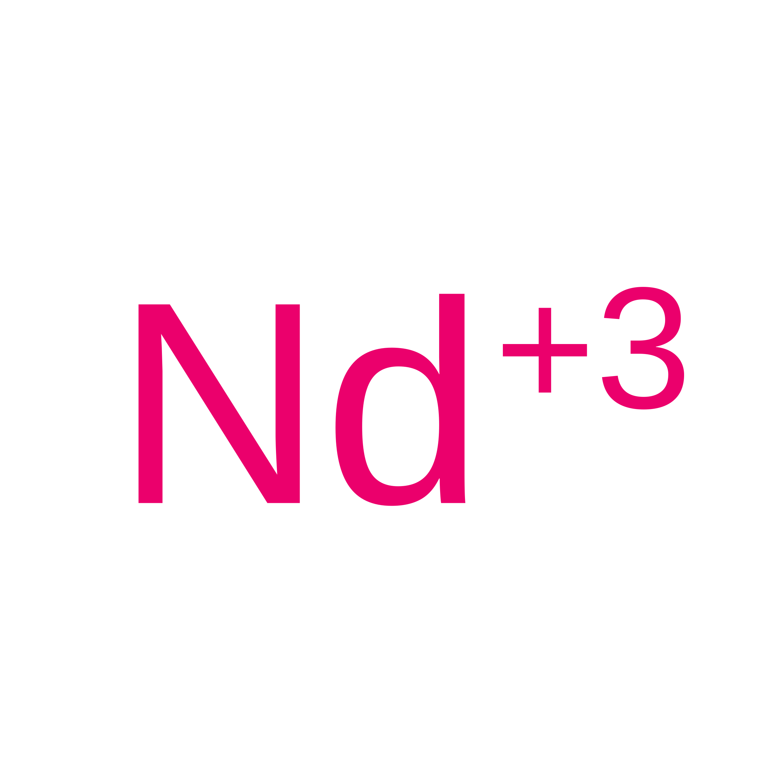
Neodymium Ion
Nd
UYIXUPGBIXNDHN-UHFFFAOYSA-N
SO4
(Subject of Investigation/LOI)

Query on SO4


Download:Ideal Coordinates CCD File
G [auth A]SULFATE ION
O4 S
QAOWNCQODCNURD-UHFFFAOYSA-L
CA
(Subject of Investigation/LOI)

Query on CA


Download:Ideal Coordinates CCD File
F [auth A],
K [auth B]
CALCIUM ION
Ca
BHPQYMZQTOCNFJ-UHFFFAOYSA-N

Experimental Data & Validation

Experimental Data

  • Method: X-RAY DIFFRACTION
  • Resolution: 0.99 Å
  • R-Value Free:  0.144 (Depositor), 0.138 (DCC) 
  • R-Value Work:  0.136 (Depositor), 0.130 (DCC) 
  • R-Value Observed: 0.136 (Depositor) 
Space Group: P 1 21 1
Unit Cell:
Length ( Å )Angle ( ˚ )
a = 40.242α = 90
b = 76.377β = 113.35
c = 42.671γ = 90
Software Package:
Software NamePurpose
PHENIXrefinement
PDB_EXTRACTdata extraction
autoPROCdata reduction
autoPROCdata scaling
PHENIXphasing

Structure Validation

View Full Validation Report


Ligand Structure Quality Assessment 


Entry History 

& Funding Information

Deposition Data


Funding OrganizationLocationGrant Number
National Institutes of Health/National Institute of General Medical Sciences (NIH/NIGMS)United States--

Revision History  (Full details and data files)

  • Version 1.0: 2026-08-19
    Type: Initial release
  • Version 1.1: 2026-09-09
    Changes: Database references
  • Version 1.2: 2026-09-16
    Changes: Database references