9PFX | pdb_00009pfx

Crystal structure of native HDO-family N-oxygenase RohS from Pseudomonas brassicacearum strain DF41 (PbrRohS)


Experimental Data Snapshot

  • Method: X-RAY DIFFRACTION
  • Resolution: 2.20 Å
  • R-Value Free: 
    0.218 (Depositor), 0.218 (DCC) 
  • R-Value Work: 
    0.183 (Depositor), 0.184 (DCC) 
  • R-Value Observed: 
    0.185 (Depositor) 

Starting Model: experimental
View more details

wwPDB Validation 3D Report Full Report

Validation slider image for 9PFX

Ligand Structure Quality Assessment 


This is version 1.1 of the entry. See complete history

Literature

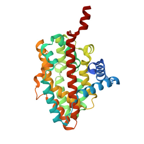
Crystal structure of RohS, a heme-oxygenase-like N-oxygenase from azomycin biosynthesis.

Wei, Z.W.Salamon, P.Higgins, M.A.Ryan, K.S.

(2026) J Biological Chem 302: 111389-111389

  • DOI: https://doi.org/10.1016/j.jbc.2026.111389
  • Primary Citation Related Structures: 
    9PFW, 9PFX

  • PubMed Abstract: 

    The nitro group is an important functional group found in the nitroimidazoles, antibiotic therapeutics for anaerobic pathogens. In the biosynthetic pathway to the nitroimidazole antibiotic azomycin, a nitro-forming enzyme RohS - a member of the heme-oxygenase-like dimetal/domain-containing oxidase/oxygenase (HDO) family - catalyzes a six-electron oxidation of 2-aminoimidazole to 2-nitroimidazole. Here we present the 2.20 Å resolution crystal structure of RohS and identify a potential active site pocket consisting of seven key residues important for metal coordination. By comparing the structures and sequences of two RohS homologs - one functionally active and one inactive - we convert the inactive RohS to its active form, thus revealing a key residue for metal coordination in RohS catalysis. Altogether, our work provides structural basis for further mechanistic investigation of this six-electron oxidation process and provides insight into the expanding repertoire of the HDO protein family and nitro-formation N-oxygenases.


  • Organizational Affiliation
    • Department of Chemistry, The University of British Columbia, Vancouver, British Columbia, Canada.

Macromolecule Content 

  • Total Structure Weight: 35.15 kDa 
  • Atom Count: 2,459 
  • Modeled Residue Count: 289 
  • Deposited Residue Count: 306 
  • Unique protein chains: 1

Macromolecules

Find similar proteins by:|  3D Structure
Entity ID: 1
MoleculeChains  Sequence LengthOrganismDetailsImage
PbrRohS306Pseudomonas brassicacearumMutation(s): 0 

Experimental Data & Validation

Experimental Data

  • Method: X-RAY DIFFRACTION
  • Resolution: 2.20 Å
  • R-Value Free:  0.218 (Depositor), 0.218 (DCC) 
  • R-Value Work:  0.183 (Depositor), 0.184 (DCC) 
  • R-Value Observed: 0.185 (Depositor) 
Space Group: P 42
Unit Cell:
Length ( Å )Angle ( ˚ )
a = 93.857α = 90
b = 93.857β = 90
c = 43.286γ = 90
Software Package:
Software NamePurpose
PHENIXrefinement
PDB_EXTRACTdata extraction
XDSdata reduction
SCALAdata scaling
PHASERphasing

Structure Validation

View Full Validation Report


Ligand Structure Quality Assessment 


Entry History 

& Funding Information

Deposition Data


Funding OrganizationLocationGrant Number
Natural Sciences and Engineering Research Council (NSERC, Canada)CanadaRGPIN-2021-02626

Revision History  (Full details and data files)

  • Version 1.0: 2026-04-01
    Type: Initial release
  • Version 1.1: 2026-05-13
    Changes: Database references