9PFM | pdb_00009pfm

Crystal structure of human 15-PGDH in complex with small molecule compound 8b


Experimental Data Snapshot

  • Method: X-RAY DIFFRACTION
  • Resolution: 1.88 Å
  • R-Value Free: 
    0.230 (Depositor), 0.232 (DCC) 
  • R-Value Work: 
    0.179 (Depositor), 0.180 (DCC) 
  • R-Value Observed: 
    0.182 (Depositor) 

Starting Model: experimental
View more details

wwPDB Validation 3D Report Full Report

Validation slider image for 9PFM

Ligand Structure Quality Assessment 


This is version 1.1 of the entry. See complete history

Literature

Knowledge and Structure-Based Drug Design of 15-PGDH Inhibitors.

Dodda, L.S.Campos, S.Ciccone, D.Carreiro, S.Leit, S.Brennan, D.Zephyr, J.Jacques-O'Hagan, S.Kumar, S.Kuo, F.S.Shaik, M.M.Price, D.J.Loh, C.Edmondson, S.D.Tummino, P.Kaila, N.

(2025) J Med Chem 68: 18436-18462

  • DOI: https://doi.org/10.1021/acs.jmedchem.5c01231
  • Primary Citation Related Structures: 
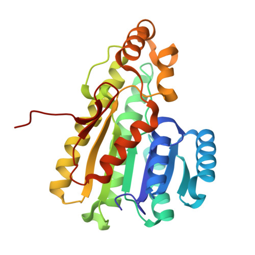
    9PFL, 9PFM

  • PubMed Abstract: 

    PGE2 plays important roles in immune cell function and in potentiating tissue regeneration. 15-PGDH is the key enzyme involved in inactivation of PGE2 and its inhibition therefore provides valuable therapeutic opportunity. We have solved the first cocrystal structure of 15-PGDH bound to small molecule inhibitors, enabling us to efficiently investigate and understand the key functionalities required for potency. Rational structure-based design coupled with a host of advanced computational methods, including FEP+ and WaterMap, were used to develop novel series of 15-PGDH inhibitors. Of note, a machine-learning (ML) model trained with potencies predicted by FEP+ yielded a powerful tool to guide synthetic priority across a large virtual chemical library. Ultimately, a lead compound demonstrated elevation of colonic PGE2 following IP administration in mice, consistent with our therapeutic hypothesis.


  • Organizational Affiliation
    • Nimbus Therapeutics, Boston, Massachusetts 02210, United States.

Macromolecule Content 

  • Total Structure Weight: 29.38 kDa 
  • Atom Count: 2,184 
  • Modeled Residue Count: 259 
  • Deposited Residue Count: 259 
  • Unique protein chains: 1

Macromolecules

Find similar proteins by:|  3D Structure
Entity ID: 1
MoleculeChains  Sequence LengthOrganismDetailsImage
15-hydroxyprostaglandin dehydrogenase [NAD(+)]259Homo sapiensMutation(s): 0 
Gene Names: HPGDPGDH1SDR36C1
EC: 1.1.1.141 (PDB Primary Data), 1.1.1 (PDB Primary Data), 1.1.1.232 (PDB Primary Data)
UniProt & NIH Common Fund Data Resources
Find proteins for P15428 (Homo sapiens)
Explore P15428 
Go to UniProtKB:  P15428
PHAROS:  P15428
GTEx:  ENSG00000164120 
Entity Groups
Sequence Clusters30% Identity50% Identity70% Identity90% Identity95% Identity100% Identity
UniProt GroupP15428
Sequence Annotations
Expand
Reference Sequence

Experimental Data & Validation

Experimental Data

  • Method: X-RAY DIFFRACTION
  • Resolution: 1.88 Å
  • R-Value Free:  0.230 (Depositor), 0.232 (DCC) 
  • R-Value Work:  0.179 (Depositor), 0.180 (DCC) 
  • R-Value Observed: 0.182 (Depositor) 
Space Group: P 61 2 2
Unit Cell:
Length ( Å )Angle ( ˚ )
a = 82.71α = 90
b = 82.71β = 90
c = 161.88γ = 120
Software Package:
Software NamePurpose
REFMACrefinement
XDSdata reduction
Aimlessdata scaling
PHASERphasing

Structure Validation

View Full Validation Report


Ligand Structure Quality Assessment 


Entry History 

& Funding Information

Deposition Data


Funding OrganizationLocationGrant Number
Not funded--

Revision History  (Full details and data files)

  • Version 1.0: 2025-09-03
    Type: Initial release
  • Version 1.1: 2025-09-17
    Changes: Database references