9PCQ | pdb_00009pcq

Phosphorylation of a Conserved Aspartate at the Eukaryotic Elongation Factor 2 Kinase Catalytic Site


Experimental Data Snapshot

  • Method: X-RAY DIFFRACTION
  • Resolution: 2.30 Å
  • R-Value Free: 
    0.243 (Depositor), 0.242 (DCC) 
  • R-Value Work: 
    0.211 (Depositor), 0.213 (DCC) 
  • R-Value Observed: 
    0.213 (Depositor) 

Starting Model: experimental
View more details

wwPDB Validation 3D Report Full Report

Validation slider image for 9PCQ

Ligand Structure Quality Assessment 


This is version 1.0 of the entry. See complete history

Literature

Phosphorylation of a conserved aspartate in the catalytic site of eukaryotic elongation factor 2 kinase.

Piserchio, A.Isiorho, E.A.Abzalimov, R.Dalby, K.N.Ghose, R.

(2026) Protein Sci 35: e70442-e70442

  • DOI: https://doi.org/10.1002/pro.70442
  • Primary Citation Related Structures: 
    9PCQ

  • PubMed Abstract: 

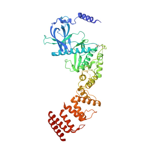
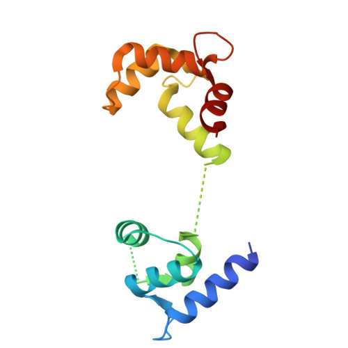
    Eukaryotic elongation factor 2 kinase (eEF-2K) is a member of the α-kinase family of atypical serine/threonine kinases. eEF-2K, the only calmodulin-activated α-kinase, phosphorylates the ribosome-associated GTPase, eukaryotic elongation factor 2 (eEF-2), suppressing translational elongation. α-kinases, including eEF-2K, possess catalytic site geometries that are distinct from those of typical kinases, suggesting possible divergence in their phospho-transfer mechanisms. Unlike typical protein kinases, where chemistry is known to proceed through a sequential mechanism involving a ternary kinase-substrate-ATP•Mg 2+ complex, the nature of the chemical step catalyzed by α-kinases remains poorly defined. Here, multiple orthogonal lines of evidence, including a crystal structure and solution-state mass spectrometry data, suggest phosphorylation of a catalytically essential aspartate residue (D284) at the eEF-2K active site. Previous crystallographic evidence of the presence of a phospho-aspartate at an equivalent position (D766) in the related Dictyostelium α-kinase MHCK-A strongly suggests that this species represents a conserved active-site feature in α-kinase family members, despite their disparate modes of activation. This observation, together with existing kinetics data on eEF-2K, raises the possibility that phospho-transfer chemistry in α-kinases occurs via an ordered stepwise mechanism involving a phospho-enzyme intermediate, contrasting with typical protein kinases.


  • Organizational Affiliation
    • Department of Chemistry and Biochemistry, The City College of New York, New York, New York, USA.

Macromolecule Content 

  • Total Structure Weight: 78.22 kDa 
  • Atom Count: 5,139 
  • Modeled Residue Count: 614 
  • Deposited Residue Count: 678 
  • Unique protein chains: 2

Macromolecules

Find similar proteins by:|  3D Structure
Entity ID: 1
MoleculeChains  Sequence LengthOrganismDetailsImage
Eukaryotic elongation factor 2 kinase531Homo sapiensMutation(s): 0 
Gene Names: EEF2K
EC: 2.7.11.20
UniProt & NIH Common Fund Data Resources
Find proteins for O00418 (Homo sapiens)
Explore O00418 
Go to UniProtKB:  O00418
PHAROS:  O00418
GTEx:  ENSG00000103319 
Entity Groups
Sequence Clusters30% Identity50% Identity70% Identity90% Identity95% Identity100% Identity
UniProt GroupO00418
Sequence Annotations
Expand
Reference Sequence
Find similar proteins by:|  3D Structure
Entity ID: 2
MoleculeChains  Sequence LengthOrganismDetailsImage
Calmodulin-1B [auth B00A]147Homo sapiensMutation(s): 0 
Gene Names: CALM1CALMCAMCAM1
UniProt & NIH Common Fund Data Resources
Find proteins for P0DP23 (Homo sapiens)
Explore P0DP23 
Go to UniProtKB:  P0DP23
PHAROS:  P0DP23
GTEx:  ENSG00000198668 
Entity Groups
Sequence Clusters30% Identity50% Identity70% Identity90% Identity95% Identity100% Identity
UniProt GroupP0DP23
Sequence Annotations
Expand
Reference Sequence

Small Molecules

Ligands 4 Unique
IDChains Name / Formula / InChI Key2D Diagram3D Interactions
ADP
(Subject of Investigation/LOI)

Query on ADP


Download:Ideal Coordinates CCD File
C [auth A],
D [auth A]
ADENOSINE-5'-DIPHOSPHATE
C10 H15 N5 O10 P2
XTWYTFMLZFPYCI-KQYNXXCUSA-N
ZN

Query on ZN


Download:Ideal Coordinates CCD File
F [auth A]ZINC ION
Zn
PTFCDOFLOPIGGS-UHFFFAOYSA-N
MN
(Subject of Investigation/LOI)

Query on MN


Download:Ideal Coordinates CCD File
E [auth A],
I [auth B00A]
MANGANESE (II) ION
Mn
WAEMQWOKJMHJLA-UHFFFAOYSA-N
CA

Query on CA


Download:Ideal Coordinates CCD File
G [auth B00A],
H [auth B00A]
CALCIUM ION
Ca
BHPQYMZQTOCNFJ-UHFFFAOYSA-N
Modified Residues  2 Unique
IDChains TypeFormula2D DiagramParent
PHD
Query on PHD
A
L-PEPTIDE LINKINGC4 H8 N O7 PASP
TPO
Query on TPO
A
L-PEPTIDE LINKINGC4 H10 N O6 PTHR

Experimental Data & Validation

Experimental Data

  • Method: X-RAY DIFFRACTION
  • Resolution: 2.30 Å
  • R-Value Free:  0.243 (Depositor), 0.242 (DCC) 
  • R-Value Work:  0.211 (Depositor), 0.213 (DCC) 
  • R-Value Observed: 0.213 (Depositor) 
Space Group: P 21 21 21
Unit Cell:
Length ( Å )Angle ( ˚ )
a = 60.464α = 90
b = 88.632β = 90
c = 154.872γ = 90
Software Package:
Software NamePurpose
PHENIXrefinement
autoPROCdata reduction
XDSdata scaling
PHASERphasing

Structure Validation

View Full Validation Report


Ligand Structure Quality Assessment 


Entry History 

& Funding Information

Deposition Data


Funding OrganizationLocationGrant Number
National Institutes of Health/National Institute of General Medical Sciences (NIH/NIGMS)United StatesR01 GM123252

Revision History  (Full details and data files)

  • Version 1.0: 2026-03-04
    Type: Initial release