9P6S | pdb_00009p6s

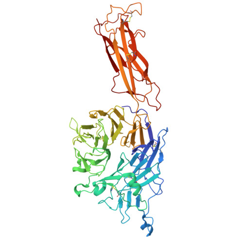
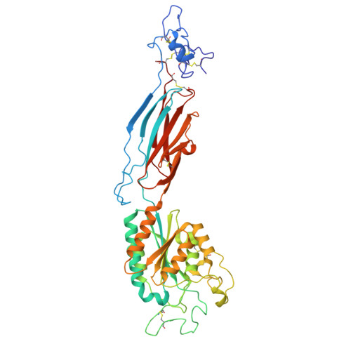
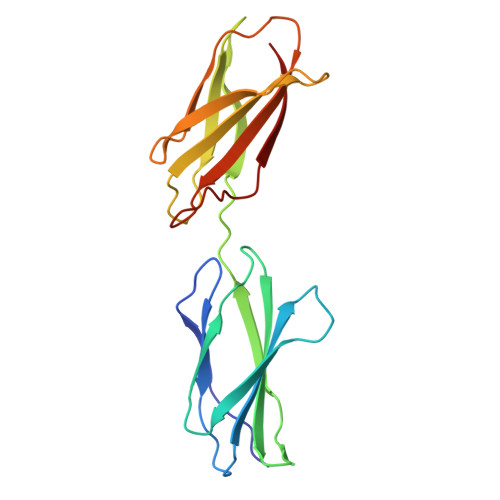
Cryo-EM structure of human integrin alpha5beta1 in complex with fibronectin (FN 7-10)


Experimental Data Snapshot

  • Method: ELECTRON MICROSCOPY
  • Resolution: 2.61 Å
  • Aggregation State: PARTICLE 
  • Reconstruction Method: SINGLE PARTICLE 

Starting Models: experimental
View more details

wwPDB Validation 3D Report Full Report

Validation slider image for 9P6S

This is version 1.0 of the entry. See complete history

Literature

Allosteric regulation of fibronectin binding by the anti-beta 1 integrin antibody TS2/16.

Ding, J.Fantini, D.A.Dedden, D.Schumacher, S.Biertumpfel, C.Mizuno, N.

(2026) PNAS Nexus 5: pgag044-pgag044

  • DOI: https://doi.org/10.1093/pnasnexus/pgag044
  • Primary Citation Related Structures: 
    9P6S

  • PubMed Abstract: 

    The monoclonal antibody (mAb) TS2/16 stabilizes the active conformation of β1 integrin, enhancing its adhesive capacity on the cell surface. However, the molecular mechanism by which TS2/16 modulates integrin affinity for extracellular ligands remains unclear. Using endogenous full-length α5β1 integrin purified from human placenta, we determined the structure of integrin α5β1 with fibronectin up to 2.61-Å resolution in the absence of TS2/16, capturing the active form without its aid, and performed comparative B-factor-based analysis and CABS-Flex simulation with and without TS2/16. Despite no global conformational differences, we found that TS2/16 interacts with α2 helix of the integrin β1 subunit and contacts the C-terminus of α3 helix, leading to a localized decrease in B factor. This interaction allosterically alters the dynamics of α2-α3 loop despite not being in direct contact with TS2/16. Notably, this loop directly engages fibronectin, and its dynamic change underlies the enhanced ligand-binding affinity and explains increased cell adhesion observed with TS2/16. These findings reveal an allosteric mechanism of integrin regulation by TS2/16 and offer insights for the rational design of therapeutic antibodies targeting integrin-mediated adhesion in pathological contexts such as inflammation and cancer.


  • Organizational Affiliation
    • Laboratory of Structural Cell Biology, National Heart, Lung, and Blood Institute, National Institutes of Health, 50 South Dr, Bethesda, MD 20892, USA.

Macromolecule Content 

  • Total Structure Weight: 139.28 kDa 
  • Atom Count: 9,778 
  • Modeled Residue Count: 1,224 
  • Deposited Residue Count: 1,224 
  • Unique protein chains: 3

Macromolecules

Find similar proteins by:|  3D Structure
Entity ID: 1
MoleculeChains  Sequence LengthOrganismDetailsImage
Integrin alpha-5601Homo sapiensMutation(s): 0 
UniProt & NIH Common Fund Data Resources
Find proteins for P08648 (Homo sapiens)
Explore P08648 
Go to UniProtKB:  P08648
PHAROS:  P08648
GTEx:  ENSG00000161638 
Entity Groups
Sequence Clusters30% Identity50% Identity70% Identity90% Identity95% Identity100% Identity
UniProt GroupP08648
Glycosylation
Glycosylation Sites: 6Go to GlyGen: P08648-1
Sequence Annotations
Expand
Reference Sequence
Find similar proteins by:|  3D Structure
Entity ID: 2
MoleculeChains  Sequence LengthOrganismDetailsImage
Integrin beta-1440Homo sapiensMutation(s): 0 
UniProt & NIH Common Fund Data Resources
Find proteins for P05556 (Homo sapiens)
Explore P05556 
Go to UniProtKB:  P05556
PHAROS:  P05556
GTEx:  ENSG00000150093 
Entity Groups
Sequence Clusters30% Identity50% Identity70% Identity90% Identity95% Identity100% Identity
UniProt GroupP05556
Glycosylation
Glycosylation Sites: 5Go to GlyGen: P05556-1
Sequence Annotations
Expand
Reference Sequence
Find similar proteins by:|  3D Structure
Entity ID: 3
MoleculeChains  Sequence LengthOrganismDetailsImage
Fibronectin183Homo sapiensMutation(s): 0 
Gene Names: FN1FN
UniProt & NIH Common Fund Data Resources
Find proteins for P02751 (Homo sapiens)
Explore P02751 
Go to UniProtKB:  P02751
PHAROS:  P02751
GTEx:  ENSG00000115414 
Entity Groups
Sequence Clusters30% Identity50% Identity70% Identity90% Identity95% Identity100% Identity
UniProt GroupP02751
Sequence Annotations
Expand
Reference Sequence

Oligosaccharides

Help  
Entity ID: 4
MoleculeChains Length2D Diagram GlycosylationD Interactions
beta-D-mannopyranose-(1-4)-2-acetamido-2-deoxy-beta-D-glucopyranose-(1-4)-2-acetamido-2-deoxy-beta-D-glucopyranose
D, G, J
3N-Glycosylation
Glycosylation Resources
GlyTouCan: G15407YE
GlyCosmos: G15407YE
GlyGen: G15407YE
Entity ID: 5
MoleculeChains Length2D Diagram GlycosylationD Interactions
alpha-D-mannopyranose-(1-6)-beta-D-mannopyranose-(1-4)-2-acetamido-2-deoxy-beta-D-glucopyranose-(1-4)-2-acetamido-2-deoxy-beta-D-glucopyranose
E
4N-Glycosylation
Glycosylation Resources
GlyTouCan: G22573RC
GlyCosmos: G22573RC
GlyGen: G22573RC
Entity ID: 6
MoleculeChains Length2D Diagram GlycosylationD Interactions
2-acetamido-2-deoxy-beta-D-glucopyranose-(1-2)-alpha-D-mannopyranose-(1-6)-[alpha-D-mannopyranose-(1-3)]beta-D-mannopyranose-(1-4)-2-acetamido-2-deoxy-beta-D-glucopyranose-(1-4)-2-acetamido-2-deoxy-beta-D-glucopyranose
F
6N-Glycosylation
Glycosylation Resources
GlyTouCan: G64481DJ
GlyCosmos: G64481DJ
GlyGen: G64481DJ
Entity ID: 7
MoleculeChains Length2D Diagram GlycosylationD Interactions
2-acetamido-2-deoxy-beta-D-glucopyranose-(1-4)-2-acetamido-2-deoxy-beta-D-glucopyranose
H, I
2N-Glycosylation
Glycosylation Resources
GlyTouCan: G42666HT
GlyCosmos: G42666HT
GlyGen: G42666HT

Small Molecules

Ligands 2 Unique
IDChains Name / Formula / InChI Key2D Diagram3D Interactions
NAG

Query on NAG


Download:Ideal Coordinates CCD File
O [auth B],
P [auth B],
Q [auth B],
R [auth B]
2-acetamido-2-deoxy-beta-D-glucopyranose
C8 H15 N O6
OVRNDRQMDRJTHS-FMDGEEDCSA-N
MN

Query on MN


Download:Ideal Coordinates CCD File
K [auth A]
L [auth A]
M [auth A]
N [auth A]
S [auth B]
K [auth A],
L [auth A],
M [auth A],
N [auth A],
S [auth B],
T [auth B],
U [auth B]
MANGANESE (II) ION
Mn
WAEMQWOKJMHJLA-UHFFFAOYSA-N

Experimental Data & Validation

Experimental Data

  • Method: ELECTRON MICROSCOPY
  • Resolution: 2.61 Å
  • Aggregation State: PARTICLE 
  • Reconstruction Method: SINGLE PARTICLE 
EM Software:
TaskSoftware PackageVersion
RECONSTRUCTIONRELION3.0
MODEL REFINEMENTCoot
MODEL REFINEMENTPHENIX1.21_5207

Structure Validation

View Full Validation Report


Entry History 

& Funding Information

Deposition Data


Funding OrganizationLocationGrant Number
National Institutes of Health/National Heart, Lung, and Blood Institute (NIH/NHLBI)United States1ZIAHL006264

Revision History  (Full details and data files)

  • Version 1.0: 2026-03-25
    Type: Initial release