9P6C | pdb_00009p6c

RTX domain block V of adenylate cyclase toxin with mutations D1533N, A1542N, D1560N, S1569N, D1587N, H1598N, H1608N

  • Classification: TOXIN
  • Organism(s): Bordetella pertussis
  • Expression System: Escherichia coli
  • Mutation(s): Yes 

  • Deposited: 2025-06-18 Released: 2025-12-24 
  • Deposition Author(s): Chang, M.P., Mai, D.J., Gudinas, A.P., Fernandez, D.
  • Funding Organization(s): Department of Defense (DOD, United States), National Institutes of Health/National Institute of General Medical Sciences (NIH/NIGMS), Department of Energy (DOE, United States), National Science Foundation (NSF, United States)

Experimental Data Snapshot

  • Method: X-RAY DIFFRACTION
  • Resolution: 1.99 Å
  • R-Value Free: 
    0.292 (Depositor), 0.293 (DCC) 
  • R-Value Work: 
    0.228 (Depositor), 0.229 (DCC) 
  • R-Value Observed: 
    0.232 (Depositor) 

Starting Model: experimental
View more details

wwPDB Validation   3D Report Full Report


This is version 1.0 of the entry. See complete history


Literature

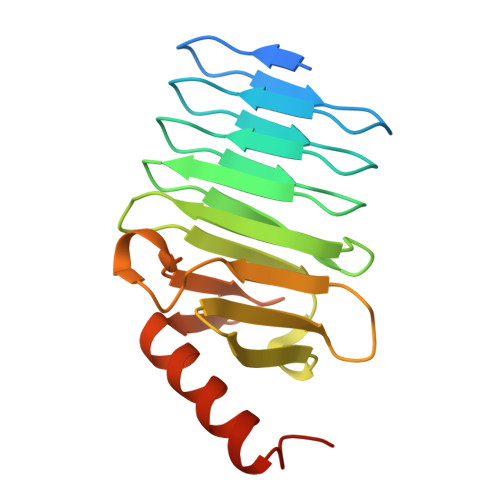
Repetitive proteins that undergo large conformational changes evade structural prediction algorithms.

Chang, M.P.Jin, T.Gudinas, A.P.Fernandez, D.Alexander-Katz, A.Matsui, T.Mai, D.J.

(2025) J Chem Phys 163

  • DOI: https://doi.org/10.1063/5.0304777
  • Primary Citation of Related Structures:  
    9P6C

  • PubMed Abstract: 

    Protein structure prediction algorithms, such as AlphaFold, have accelerated protein design and advanced the understanding of the relationship between amino acid sequence and protein structure. However, these algorithms are limited in their ability to predict the structures of conformationally dynamic, intrinsically disordered, and stimuli-responsive proteins. To evaluate sequence-to-structure predictions of such challenging proteins, we explored a class of conformationally dynamic, repeats-in-toxin (RTX) proteins. RTX proteins adopt intrinsically disordered conformations in the absence of calcium and undergo reversible folding into β-roll structures upon binding to calcium. RTX proteins are characterized by tandem repeats of the sequence GGXGXDXUX, in which X can be any amino acid and U is an aliphatic amino acid. We designed RTX sequence variants with global substitutions of nonconserved amino acids, tandem repeats of consensus sequences GGAGXDTLY, and tandem repeats of scrambled sequences GGAGXDTYL. AlphaFold2 and AlphaFold3 predicted that all of these RTX variants adopt β-roll structures, characteristic of wild-type RTX bound to calcium. However, modeling the predicted structures with molecular dynamics simulations and characterizing the protein variants with circular dichroism spectroscopy, small-angle x-ray scattering, and x-ray crystallography revealed that variants adopt diverse, sequence-dependent structures in the absence and presence of calcium. To better design proteins for applications in biotechnology and sustainability, it is critical to build predictive tools that consider intrinsically disordered protein states and validate these tools with multi-mode, multi-scale experimental data.


  • Organizational Affiliation
    • Department of Materials Science and Engineering, Stanford University, Stanford, California 94305, USA.

Macromolecules
Find similar proteins by:  (by identity cutoff)  |  3D Structure
Entity ID: 1
MoleculeChains Sequence LengthOrganismDetailsImage
Hemolysin
A, B
180Bordetella pertussisMutation(s): 7 
Gene Names: cyacyaABP0760
UniProt
Find proteins for P0DKX7 (Bordetella pertussis (strain Tohama I / ATCC BAA-589 / NCTC 13251))
Explore P0DKX7 
Go to UniProtKB:  P0DKX7
Entity Groups  
Sequence Clusters30% Identity50% Identity70% Identity90% Identity95% Identity100% Identity
UniProt GroupP0DKX7
Sequence Annotations
Expand
  • Reference Sequence
Small Molecules
Ligands 1 Unique
IDChains Name / Formula / InChI Key2D Diagram3D Interactions
CA (Subject of Investigation/LOI)
Query on CA

Download Ideal Coordinates CCD File 
C [auth A]
D [auth A]
E [auth A]
F [auth A]
G [auth A]
C [auth A],
D [auth A],
E [auth A],
F [auth A],
G [auth A],
H [auth A],
I [auth A],
J [auth A],
K [auth B],
L [auth B],
M [auth B],
N [auth B],
O [auth B],
P [auth B],
Q [auth B],
R [auth B]
CALCIUM ION
Ca
BHPQYMZQTOCNFJ-UHFFFAOYSA-N
Experimental Data & Validation

Experimental Data

  • Method: X-RAY DIFFRACTION
  • Resolution: 1.99 Å
  • R-Value Free:  0.292 (Depositor), 0.293 (DCC) 
  • R-Value Work:  0.228 (Depositor), 0.229 (DCC) 
  • R-Value Observed: 0.232 (Depositor) 
Space Group: P 1
Unit Cell:
Length ( Å )Angle ( ˚ )
a = 33.305α = 84.13
b = 38.003β = 81.47
c = 57.016γ = 67.61
Software Package:
Software NamePurpose
REFMACrefinement
PDB_EXTRACTdata extraction
XDSdata reduction
SCALAdata scaling
PHASERphasing

Structure Validation

View Full Validation Report


Entry History & Funding Information

Deposition Data


Funding OrganizationLocationGrant Number
Department of Defense (DOD, United States)United StatesFA9550-22-1-0241
National Institutes of Health/National Institute of General Medical Sciences (NIH/NIGMS)United StatesP30GM133894
Department of Energy (DOE, United States)United StatesDE-AC02-76SF00515
National Science Foundation (NSF, United States)United StatesDGE-1656518

Revision History  (Full details and data files)

  • Version 1.0: 2025-12-24
    Type: Initial release