


Structure of phospholipase D BetaIB1i from Sicarius terrosus venom, H47N mutant bound to product and substrate sphingolipids at 1.85 A resolution
Sundman, A.K., Montfort, W.R., Binford, G.J., Cordes, M.H.To be published.
Experimental Data Snapshot
Starting Model: experimental
View more details
Entity ID: 1 | |||||
|---|---|---|---|---|---|
| Molecule | Chains | Sequence Length | Organism | Details | Image |
| Dermonecrotic toxin StSicTox-betaIB1i | 301 | Sicarius terrosus | Mutation(s): 1 EC: 4.6.1 | 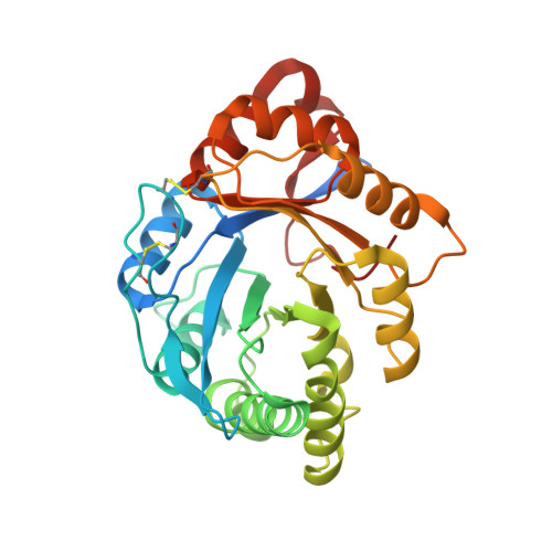 | |
UniProt | |||||
Entity Groups | |||||
| Sequence Clusters | 30% Identity50% Identity70% Identity90% Identity95% Identity100% Identity | ||||
| UniProt Group | A0A0D4WV12 | ||||
Sequence AnnotationsExpand | |||||
Reference Sequence | |||||
| Ligands 4 Unique | |||||
|---|---|---|---|---|---|
| ID | Chains | Name / Formula / InChI Key | 2D Diagram | 3D Interactions | |
| A1A43 (Subject of Investigation/LOI) Download:Ideal Coordinates CCD File | C [auth A] D [auth A] E [auth A] J [auth B] K [auth B] | 2-aminoethyl (2S,3R,4E)-2-dodecanamido-3-hydroxyheptadec-4-en-1-yl hydrogen (S)-phosphate C31 H63 N2 O6 P ZIEWVPJUBVRCAL-DQBIDPIGSA-N |  | ||
| MPD Download:Ideal Coordinates CCD File | H [auth A], I [auth A] | (4S)-2-METHYL-2,4-PENTANEDIOL C6 H14 O2 SVTBMSDMJJWYQN-YFKPBYRVSA-N |  | ||
| MG Download:Ideal Coordinates CCD File | G [auth A], N [auth B] | MAGNESIUM ION Mg JLVVSXFLKOJNIY-UHFFFAOYSA-N |  | ||
| NA Download:Ideal Coordinates CCD File | F [auth A], M [auth B] | SODIUM ION Na FKNQFGJONOIPTF-UHFFFAOYSA-N |  | ||
| Length ( Å ) | Angle ( ˚ ) |
|---|---|
| a = 80.789 | α = 90 |
| b = 105.854 | β = 93.62 |
| c = 109.108 | γ = 90 |
| Software Name | Purpose |
|---|---|
| REFMAC | refinement |
| PDB_EXTRACT | data extraction |
| PROTEUM2 | data reduction |
| Aimless | data scaling |
| MOLREP | phasing |
| Funding Organization | Location | Grant Number |
|---|---|---|
| National Science Foundation (NSF, United States) | United States | CHE-1808716 |


