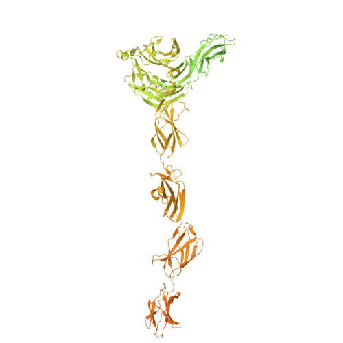
Structural insights into the activation of the chicken ROS1 receptor by the NEL/NICOL ligand complex.
An, W., Zhang, X., Bai, X.C.(2026) Nat Commun 17
- PubMed: 41735298 Search on PubMed
- DOI: https://doi.org/10.1038/s41467-026-69942-8
- Primary Citation Related Structures:
9OYZ, 9OZ1, 9OZ6, 9OZ8, 9OZC, 9OZH, 9OZI - PubMed Abstract:
The receptor tyrosine kinase ROS1 plays essential roles in cell growth and sperm maturation, yet its activation mechanism has remained poorly understood. Here, we report high-resolution cryo-electron microscopy (cryo-EM) structures of chicken ROS1 in its ligand-free form, in complex with its ligand NEL, and with the ligand/co-ligand complex NEL/NICOL. Unliganded ROS1 adopts an arc-shaped conformation. The interaction between NEL and ROS1 is mediated by the VWC2 domain of NEL and the β1 domain of ROS1. Binding of NICOL to the coiled-coil domain of NEL stabilizes NEL into a batwing-shaped asymmetric dimer, which can recruit only one ROS1 molecule due to steric hindrance. Structural analyses and biochemical results suggest that the 2:1 NEL/NICOL complexes further oligomerize through LamG-VWC4 domain interactions, facilitating the clustering of multiple ROS1 for its activation. Functional assays confirm that both NICOL and the multimerization of NEL/NICOL complexes are required for robust ROS1 signaling. Our findings establish NICOL as a critical co-ligand for ROS1 and suggest a distinct ligand-driven oligomerization mechanism for ROS1 activation.
- Department of Biophysics, University of Texas Southwestern Medical Center, Dallas, TX, USA.
Organizational Affiliation: