9ON6 | pdb_00009on6

The pre-open state 2 structure of human P2X2 receptor channel in lipid nanodiscs with free ATP and sodium

  • Classification: MEMBRANE PROTEIN
  • Organism(s): Homo sapiens
  • Expression System: Homo sapiens
  • Mutation(s): No 

  • Deposited: 2025-05-14 Released: 2026-03-18 
  • Deposition Author(s): Dhingra, S., Swartz, K.J.
  • Funding Organization(s): National Institutes of Health/National Institute of Neurological Disorders and Stroke (NIH/NINDS)

Experimental Data Snapshot

  • Method: ELECTRON MICROSCOPY
  • Resolution: 2.60 Å
  • Aggregation State: PARTICLE 
  • Reconstruction Method: SINGLE PARTICLE 

wwPDB Validation 3D Report Full Report

Validation slider image for 9ON6

This is version 1.1 of the entry. See complete history

Literature

Mechanisms of ligand recognition and channel opening for P2X2 receptors in lipid nanodiscs.

Dhingra, S.Moog, M.Swartz, K.J.

(2026) Sci Adv 12: eaee4242-eaee4242

  • DOI: https://doi.org/10.1126/sciadv.aee4242
  • Primary Citation Related Structures: 
    9OGH, 9OHK, 9OIR, 9OJK, 9OM0, 9OMR, 9ON5, 9ON6, 9Z32

  • PubMed Abstract: 

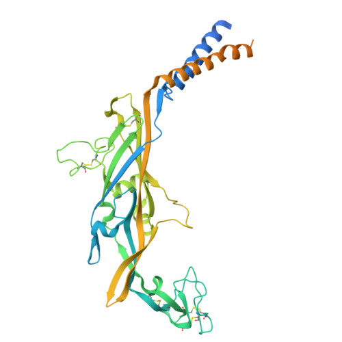
    Extracellular adenosine 5'-triphosphate (ATP) activates P2X receptor channels (P2XRs) that serve important roles in the immune and nervous systems. Available structures of P2XRs in detergents reveal that ATP binding to the extracellular domain leads to severing of subunit interfaces within transmembrane regions as the pore opens. Here we report cryo-electron microscopy structures of the human P2X2R in lipid nanodiscs in an apo closed state, with ATP 4- , Mg-ATP 2- , and suramin bound. We find that a unique Arg residue interacts with the γ-PO 4 of ATP 4- in P2X2R and underlies the requirement of this subtype for ATP 4- . Channel opening and desensitization occur when ATP 4- binds, whereas the channel remains closed when Mg-ATP 2- binds. A continuous belt of partially resolved lipids in the outer leaflet stabilizes the closed state, and the presence of lipids prevents the severing of subunit interfaces as the channel opens. These findings establish key mechanistic principles of gating for P2X2R in a membrane-like environment, providing a framework for future mechanistic studies and therapeutic development.


  • Organizational Affiliation
    • Molecular Physiology and Biophysics Section, Porter Neuroscience Research Center, National Institute of Neurological Disorders and Stroke, National Institutes of Health, Bethesda, MD 20892, USA.

Macromolecule Content 

  • Total Structure Weight: 158.96 kDa 
  • Atom Count: 7,791 
  • Modeled Residue Count: 969 
  • Deposited Residue Count: 1,413 
  • Unique protein chains: 1

Macromolecules

Find similar proteins by:|  3D Structure
Entity ID: 1
MoleculeChains  Sequence LengthOrganismDetailsImage
P2X purinoceptor 2A,
B [auth C],
C [auth B]
471Homo sapiensMutation(s): 0 
Gene Names: P2RX2P2X2
UniProt & NIH Common Fund Data Resources
Find proteins for Q9UBL9 (Homo sapiens)
Explore Q9UBL9 
Go to UniProtKB:  Q9UBL9
PHAROS:  Q9UBL9
GTEx:  ENSG00000187848 
Entity Groups
Sequence Clusters30% Identity50% Identity70% Identity90% Identity95% Identity100% Identity
UniProt GroupQ9UBL9
Glycosylation
Glycosylation Sites: 3Go to GlyGen: Q9UBL9-1
Sequence Annotations
Expand
Reference Sequence

Small Molecules

Ligands 2 Unique
IDChains Name / Formula / InChI Key2D Diagram3D Interactions
ATP
(Subject of Investigation/LOI)

Query on ATP


Download:Ideal Coordinates CCD File
G [auth A],
K [auth C],
O [auth B]
ADENOSINE-5'-TRIPHOSPHATE
C10 H16 N5 O13 P3
ZKHQWZAMYRWXGA-KQYNXXCUSA-N
NAG
(Subject of Investigation/LOI)

Query on NAG


Download:Ideal Coordinates CCD File
D [auth A]
E [auth A]
F [auth A]
H [auth C]
I [auth C]
D [auth A],
E [auth A],
F [auth A],
H [auth C],
I [auth C],
J [auth C],
L [auth B],
M [auth B],
N [auth B]
2-acetamido-2-deoxy-beta-D-glucopyranose
C8 H15 N O6
OVRNDRQMDRJTHS-FMDGEEDCSA-N

Experimental Data & Validation

Experimental Data

  • Method: ELECTRON MICROSCOPY
  • Resolution: 2.60 Å
  • Aggregation State: PARTICLE 
  • Reconstruction Method: SINGLE PARTICLE 
EM Software:
TaskSoftware PackageVersion
MODEL REFINEMENTPHENIX1.20.1_4487:
RECONSTRUCTIONcryoSPARC

Structure Validation

View Full Validation Report


Entry History 

& Funding Information

Deposition Data


Funding OrganizationLocationGrant Number
National Institutes of Health/National Institute of Neurological Disorders and Stroke (NIH/NINDS)United StatesNS003018

Revision History  (Full details and data files)

  • Version 1.0: 2026-03-18
    Type: Initial release
  • Version 1.1: 2026-06-10
    Changes: Database references