Download Mendeley
Bacterial 2',3'-cGAMP activates a SAVED effector to form membrane-disrupting filaments and restrict phage replication.
Tak, U., Schinkel, K., Walth, P., Tay, J.W., Hartwick, E.W., Whiteley, A.T.(2026) Cell Host Microbe 34: 720
- PubMed: 41903528 Search on PubMedSearch on PubMed Central
- DOI: https://doi.org/10.1016/j.chom.2026.03.004
- Primary Citation Related Structures:
9OM7 - PubMed Abstract:
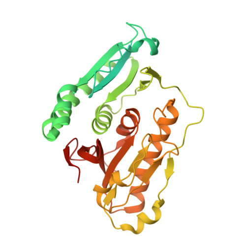
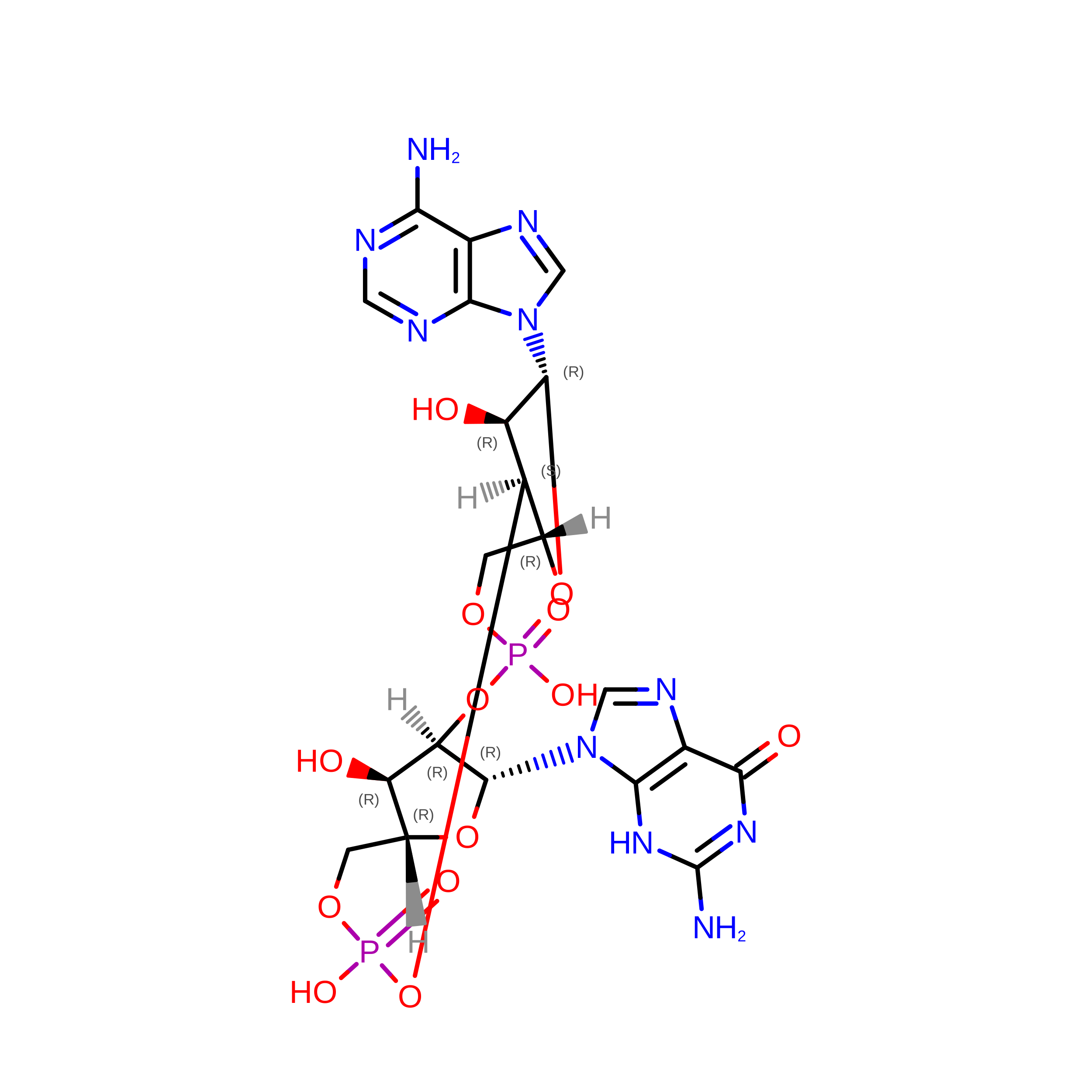
Mammalian cells initiate antiviral signaling when cyclic GMP-AMP synthase (cGAS) detects cytoplasmic DNA and synthesizes 2',3'-cyclic GMP-AMP (2',3'-cGAMP), which activates stimulator of interferon genes (STING). Similarly, bacteria use cyclic oligonucleotide-based antiphage signaling systems (CBASS) to detect phage using ancestral cGAS/DncV-like nucleotidyltransferases (CD-NTases), but they are not known to use 2',3'-cGAMP. Here, we discover a bacterial CD-NTase that produces 2',3'-cGAMP to activate a Saf-2TM-SMODS-associated fused to various effector domains (SAVED) effector (CD-NTase-associated protein 14 [Cap14]), which initiates membrane disruption to restrict phage replication. Cryo-electron microscopy (cryo-EM) reveals that Cap14 binds 2',3'-cGAMP to form a filament, while electrophysiology suggests that cGAMP activates membrane disruption. Swapping the Cap14 transmembrane domain with a nuclease domain yields a functional chimera that exclusively responds to 2',3'-cGAMP. We hypothesize that other predicted transmembrane effectors in CBASS operons disrupt membranes, and we confirm this by showing that bacterial STING homologs with transmembrane domains restrict phage through membrane disruption. These findings expand our understanding of cGAS-STING-like pathways in bacterial immunity.
- Department of Biochemistry, University of Colorado Boulder, Boulder, CO, USA.
Organizational Affiliation: