9OL6 | pdb_00009ol6

Rabbit Ryanodine Receptor 1: DMSO Control Closed Conformation


Experimental Data Snapshot

  • Method: ELECTRON MICROSCOPY
  • Resolution: 3.11 Å
  • Aggregation State: PARTICLE 
  • Reconstruction Method: SINGLE PARTICLE 

Starting Model: experimental
View more details

wwPDB Validation   3D Report Full Report


This is version 2.0 of the entry. See complete history


Literature

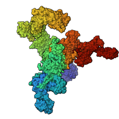
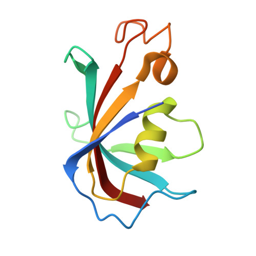
Cryo-electron microscopy reveals sequential binding and activation of Ryanodine Receptors by statin triplets.

Molinarolo, S.Valdivia, C.R.Valdivia, H.H.Van Petegem, F.

(2025) Nat Commun 16: 11508-11508

  • DOI: https://doi.org/10.1038/s41467-025-66522-0
  • Primary Citation of Related Structures:  
    9OL6, 9PWO

  • PubMed Abstract: 

    Statins are the most prescribed class of drugs and inhibit a key enzyme in the cholesterol biosynthesis pathway. Many patients have reported mild to severe muscle related symptoms and a subset are at risk for rhabdomyolysis. Sequence variants in RyR1, the skeletal muscle Ryanodine Receptor, correlate with intolerance to statins, but whether RyR1 can bind statins directly has remained unclear. Here we report cryo-EM structures of RyR1 in the absence and presence of atorvastatin, firmly establishing RyR1 as an unintended off-target. Our results show an unusual binding mode whereby three atorvastatin molecules bind together in a cleft formed by the pseudo-voltage sensing domain, making extensive interactions with each other and with RyR1. Atorvastatin activates RyR1 in a sequential way, whereby one statin per subunit can bind to the transmembrane region of a closed RyR1, with small structural perturbations that prime the channel for opening. Binding of two additional statins per subunit is associated with a widening of the pseudo-voltage sensing domain that triggers opening of the pore. Comparison with atorvastatin binding to HMG-CoA reductase, its intended target, offers clues on how to modify the statin to reduce RyR1 binding, while leaving binding to HMG-CoA reductase unperturbed.


  • Organizational Affiliation
    • Department of Biochemistry and Molecular Biology, The Life Sciences Institute, University of British Columbia, Vancouver, British Columbia, Canada.

Macromolecules
Find similar proteins by:  (by identity cutoff)  |  3D Structure
Entity ID: 1
MoleculeChains Sequence LengthOrganismDetailsImage
Ryanodine receptor 1A,
C [auth G],
E [auth M],
G [auth S]
5,037Oryctolagus cuniculusMutation(s): 0 
UniProt
Find proteins for P11716 (Oryctolagus cuniculus)
Explore P11716 
Go to UniProtKB:  P11716
Entity Groups  
Sequence Clusters30% Identity50% Identity70% Identity90% Identity95% Identity100% Identity
UniProt GroupP11716
Sequence Annotations
Expand
  • Reference Sequence
Find similar proteins by:  (by identity cutoff)  |  3D Structure
Entity ID: 2
MoleculeChains Sequence LengthOrganismDetailsImage
Peptidyl-prolyl cis-trans isomerase FKBP1BB,
D [auth H],
F [auth N],
H [auth T]
111Homo sapiensMutation(s): 0 
Gene Names: FKBP1BFKBP12.6FKBP1LFKBP9OTK4
EC: 5.2.1.8
UniProt & NIH Common Fund Data Resources
Find proteins for P68106 (Homo sapiens)
Explore P68106 
Go to UniProtKB:  P68106
PHAROS:  P68106
GTEx:  ENSG00000119782 
Entity Groups  
Sequence Clusters30% Identity50% Identity70% Identity90% Identity95% Identity100% Identity
UniProt GroupP68106
Sequence Annotations
Expand
  • Reference Sequence
Experimental Data & Validation

Experimental Data

  • Method: ELECTRON MICROSCOPY
  • Resolution: 3.11 Å
  • Aggregation State: PARTICLE 
  • Reconstruction Method: SINGLE PARTICLE 
EM Software:
TaskSoftware PackageVersion
MODEL REFINEMENTPHENIX1.21.1
MODEL REFINEMENTISOLDE1.7.1
RECONSTRUCTIONcryoSPARC4.4.1

Structure Validation

View Full Validation Report


Entry History & Funding Information

Deposition Data


Funding OrganizationLocationGrant Number
Canadian Institutes of Health Research (CIHR)Canada--

Revision History  (Full details and data files)

  • Version 1.0: 2025-12-03
    Type: Initial release
  • Version 1.1: 2026-01-14
    Changes: Data collection, Database references
  • Version 2.0: 2026-01-21
    Changes: Advisory, Atomic model, Data collection, Database references, Derived calculations, Structure summary