9OJV | pdb_00009ojv

Crystal structure of monoclonal J2 antibody bound to dsRNA


Experimental Data Snapshot

  • Method: X-RAY DIFFRACTION
  • Resolution: 2.85 Å
  • R-Value Free: 
    0.262 (Depositor), 0.262 (DCC) 
  • R-Value Work: 
    0.212 (Depositor), 0.212 (DCC) 
  • R-Value Observed: 
    0.214 (Depositor) 

Starting Model: experimental
View more details

wwPDB Validation 3D Report Full Report

Validation slider image for 9OJV

Ligand Structure Quality Assessment 


This is version 1.1 of the entry. See complete history

Literature

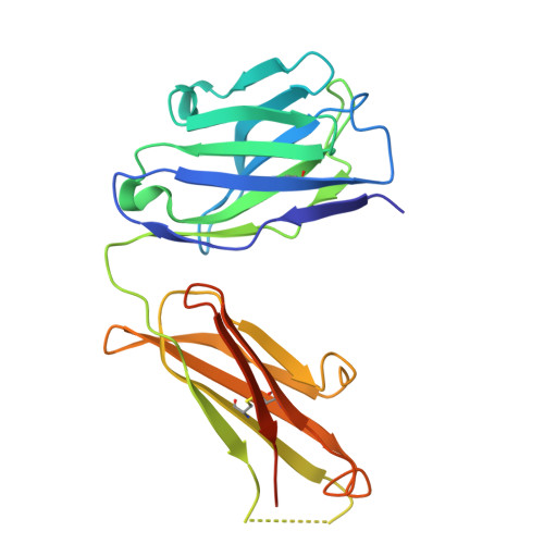
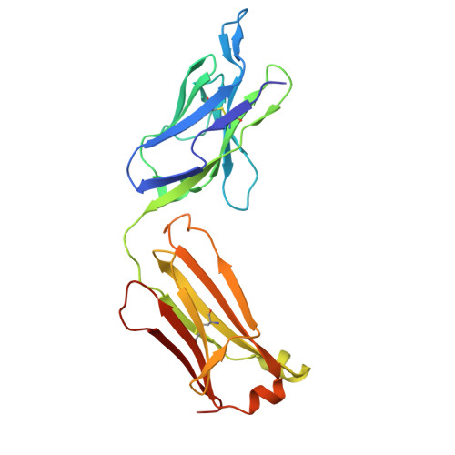
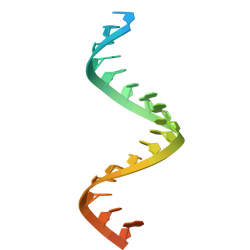
Structural basis of double-stranded RNA recognition by the J2 monoclonal antibody.

Bou-Nader, C.Juma, K.M.Bothra, A.Brasington, A.J.Ghirlando, R.Suzuki, M.Garboczi, D.N.Leppla, S.H.Zhang, J.

(2025) Nat Commun 17: 635-635

  • DOI: https://doi.org/10.1038/s41467-025-67414-z
  • Primary Citation Related Structures: 
    9OJV

  • PubMed Abstract: 

    Double-stranded (ds) RNAs are major structural components of the transcriptome, hallmarks of viral infection, and primary triggers of innate immune responses. The J2 monoclonal antibody is the gold-standard method to discover and map endogenous dsRNAs across subcellular locations and cell surfaces, detect exogenous RNAs in viral infection, and surveil mRNA prophylactics and therapeutics for inflammatory dsRNAs. To define its epitope, specificity, and mechanism, we determine a 2.85 Å co-crystal structure of J2 antigen-binding fragment (Fab) bound to dsRNA. J2 uses its heavy and light chains in tandem to track the dsRNA minor groove, recognizing a staggered 8-bp duplex. J2 is highly selective for dsRNAs, requires 14 bp for robust binding, and exhibits greatly diminished binding for GC-rich dsRNAs. J2 and the R-loop-specific S9.6 antibody share a common recognition strategy distinct from intracellular dsRNA-binding proteins. This study provides mechanistic insights into dsRNA recognition and establishes a framework for reliable application and data interpretation of the J2 antibody in RNA discovery.


  • Organizational Affiliation
    • Laboratory of Molecular Biology, National Institute of Diabetes and Digestive and Kidney Diseases, Bethesda, MD, USA.

Macromolecule Content 

  • Total Structure Weight: 113.56 kDa 
  • Atom Count: 7,309 
  • Modeled Residue Count: 895 
  • Deposited Residue Count: 946 
  • Unique protein chains: 2
  • Unique nucleic acid chains: 2

Macromolecules


Find similar proteins by:|  3D Structure
Entity ID: 1
MoleculeChains  Sequence LengthOrganismDetailsImage
J2 Fab heavy chainA [auth C],
C [auth A]
230Mus musculusMutation(s): 0 
Find similar proteins by:|  3D Structure
Entity ID: 2
MoleculeChains  Sequence LengthOrganismDetailsImage
J2 Fab light chainB [auth D],
D [auth B]
219Mus musculusMutation(s): 0 
Find similar nucleic acids by:  Sequence
Entity ID: 3
MoleculeChains LengthOrganismImage
RNA strand124synthetic construct
Sequence Annotations
Expand
Reference Sequence
Find similar nucleic acids by:  Sequence
Entity ID: 4
MoleculeChains LengthOrganismImage
RNA strand224synthetic construct
Sequence Annotations
Expand
Reference Sequence

Experimental Data & Validation

Experimental Data

  • Method: X-RAY DIFFRACTION
  • Resolution: 2.85 Å
  • R-Value Free:  0.262 (Depositor), 0.262 (DCC) 
  • R-Value Work:  0.212 (Depositor), 0.212 (DCC) 
  • R-Value Observed: 0.214 (Depositor) 
Space Group: P 2 21 21
Unit Cell:
Length ( Å )Angle ( ˚ )
a = 65.391α = 90
b = 92.374β = 90
c = 194.663γ = 90
Software Package:
Software NamePurpose
PHENIXrefinement
XDSdata reduction
XSCALEdata scaling
PHENIXphasing

Structure Validation

View Full Validation Report


Ligand Structure Quality Assessment 


Entry History 

& Funding Information

Deposition Data


Funding OrganizationLocationGrant Number
National Institutes of Health/National Institute of Diabetes and Digestive and Kidney Disease (NIH/NIDDK)United States--

Revision History  (Full details and data files)

  • Version 1.0: 2025-12-10
    Type: Initial release
  • Version 1.1: 2026-03-25
    Changes: Database references