9OHU | pdb_00009ohu

CD1c presenting GM3 ganglioside


Experimental Data Snapshot

  • Method: X-RAY DIFFRACTION
  • Resolution: 2.90 Å
  • R-Value Free: 
    0.272 (Depositor), 0.268 (DCC) 
  • R-Value Work: 
    0.234 (Depositor), 0.226 (DCC) 

Starting Model: experimental
View more details

wwPDB Validation   3D Report Full Report


Ligand Structure Quality Assessment 


This is version 2.1 of the entry. See complete history


Literature

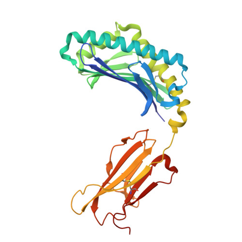
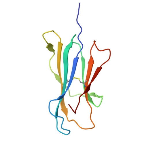
Sideways lipid presentation by the antigen-presenting molecule CD1c.

Cao, T.P.Liao, G.R.Cheng, T.Y.Chen, Y.Ciacchi, L.Fulford, T.S.Farquhar, R.Kollmorgen, J.Mayfield, J.A.Uldrich, A.P.Ng, E.Z.Q.Ogg, G.S.Godfrey, D.I.Gherardin, N.A.Chen, Y.L.Moody, D.B.Shahine, A.Rossjohn, J.

(2025) Nat Commun 

  • DOI: https://doi.org/10.1038/s41467-025-67732-2
  • Primary Citation of Related Structures:  
    9OHT, 9OHU, 9OHV, 9OHW, 9OHX, 9OHY, 9OHZ, 9OI0

  • PubMed Abstract: 

    For the MHC, MR1 and CD1 systems, antigen recognition involves contact of the membrane distal surfaces of both the αβ T cell receptor (TCR) and the antigen-presenting molecule. Whether other antigen display mechanisms by antigen-presenting molecules operate remains unknown. Here, we report mass spectrometry analyses of endogenous lipids captured by CD1c when bound to an autoreactive αβ TCR. CD1c binds twenty-six lipid species with bulky headgroups that cannot fit within the tight TCR-CD1c interface. We determined the crystal structures of CD1c presenting several gangliosides, revealing a general mechanism whereby two lipids, rather than one, are bound in the CD1c cleft. Bulky lipids are oriented sideways so that their polar headgroups protrude laterally through a side portal of the CD1c molecule - an evolutionarily conserved structural feature. The sideways-presented ganglioside headgroups do not hinder TCR binding and so represent a mechanism that allows autoreactive TCR recognition of CD1c. In addition, ex vivo studies showed that the sideways-presented gangliosides can also represent TCR recognition determinants. These findings reveal that CD1c simultaneously presents two lipid antigens from the top and side of its cleft, a general mechanism that differs markedly from other antigen-presenting molecules.


  • Organizational Affiliation
    • Infection and Immunity Program and Department of Biochemistry and Molecular Biology, Biomedicine Discovery Institute, Monash University, Clayton, VIC, Australia.

Macromolecules
Find similar proteins by:  (by identity cutoff)  |  3D Structure
Entity ID: 1
MoleculeChains Sequence LengthOrganismDetailsImage
T-cell surface glycoprotein CD1c/T-cell surface glycoprotein CD1b chimeric protein281Homo sapiensMutation(s): 0 
Gene Names: CD1C
Entity Groups  
Sequence Clusters30% Identity50% Identity70% Identity90% Identity95% Identity100% Identity
Glycosylation
Glycosylation Sites: 1
Sequence Annotations
Expand
  • Reference Sequence
Find similar proteins by:  (by identity cutoff)  |  3D Structure
Entity ID: 2
MoleculeChains Sequence LengthOrganismDetailsImage
Beta-2-microglobulin101Homo sapiensMutation(s): 0 
Gene Names: B2MCDABP0092HDCMA22P
UniProt & NIH Common Fund Data Resources
Find proteins for P61769 (Homo sapiens)
Explore P61769 
Go to UniProtKB:  P61769
PHAROS:  P61769
GTEx:  ENSG00000166710 
Entity Groups  
Sequence Clusters30% Identity50% Identity70% Identity90% Identity95% Identity100% Identity
UniProt GroupP61769
Sequence Annotations
Expand
  • Reference Sequence
Small Molecules
Ligands 6 Unique
IDChains Name / Formula / InChI Key2D Diagram3D Interactions
A1CB6 (Subject of Investigation/LOI)
Query on A1CB6

Download Ideal Coordinates CCD File 
J [auth A](2S,3R,4E)-3-hydroxy-2-octadecanamidooctadec-4-en-1-yl 5-acetamido-3,5-dideoxy-D-glycero-alpha-D-galacto-non-2-ulopyranonosyl-(2->3)-beta-D-galactopyranosyl-(1->4)-beta-D-glucopyranoside
C59 H108 N2 O21
UIKPUUZBQYTDRX-YRRBDHRDSA-N
A1CB5
Query on A1CB5

Download Ideal Coordinates CCD File 
D [auth A](2S)-2,3-dihydroxypropyl heptadecanoate
C20 H40 O4
SVUQHVRAGMNPLW-IBGZPJMESA-N
NAG
Query on NAG

Download Ideal Coordinates CCD File 
C [auth A]2-acetamido-2-deoxy-beta-D-glucopyranose
C8 H15 N O6
OVRNDRQMDRJTHS-FMDGEEDCSA-N
KZF
Query on KZF

Download Ideal Coordinates CCD File 
E [auth A]2-(cyclohexylazaniumyl)ethanesulfonate
C8 H17 N O3 S
MKWKNSIESPFAQN-UHFFFAOYSA-N
MLA
Query on MLA

Download Ideal Coordinates CCD File 
F [auth A],
G [auth A]
MALONIC ACID
C3 H4 O4
OFOBLEOULBTSOW-UHFFFAOYSA-N
CL
Query on CL

Download Ideal Coordinates CCD File 
H [auth A],
I [auth A]
CHLORIDE ION
Cl
VEXZGXHMUGYJMC-UHFFFAOYSA-M
Experimental Data & Validation

Experimental Data

  • Method: X-RAY DIFFRACTION
  • Resolution: 2.90 Å
  • R-Value Free:  0.272 (Depositor), 0.268 (DCC) 
  • R-Value Work:  0.234 (Depositor), 0.226 (DCC) 
Space Group: P 31 2 1
Unit Cell:
Length ( Å )Angle ( ˚ )
a = 88.804α = 90
b = 88.804β = 90
c = 157.198γ = 120
Software Package:
Software NamePurpose
PHENIXrefinement
Aimlessdata scaling
XDSdata reduction
PDB_EXTRACTdata extraction
PHASERphasing

Structure Validation

View Full Validation Report


Ligand Structure Quality Assessment 


Entry History & Funding Information

Deposition Data


Funding OrganizationLocationGrant Number
Australian Research Council (ARC)AustraliaDE210101031

Revision History  (Full details and data files)

  • Version 1.0: 2025-11-19
    Type: Initial release
  • Version 2.0: 2025-12-17
    Type: Coordinate replacement
    Reason: Chirality error
    Changes: Advisory, Atomic model, Data collection, Database references, Derived calculations, Polymer sequence, Refinement description, Source and taxonomy, Structure summary
  • Version 2.1: 2026-01-14
    Changes: Database references