Synthetic Rewiring of Virus-like Particles via Circular Permutation Enables Modular Peptide Display and Protein Encapsulation.
Liang, S., Butaney, K., de Castro Assumpcao, D., Jung, J., Kennedy, N.W., Tullman-Ercek, D.(2025) ACS Nano 19: 39168-39180
- PubMed: 41159643 Search on PubMed
- DOI: https://doi.org/10.1021/acsnano.5c12293
- Primary Citation Related Structures:
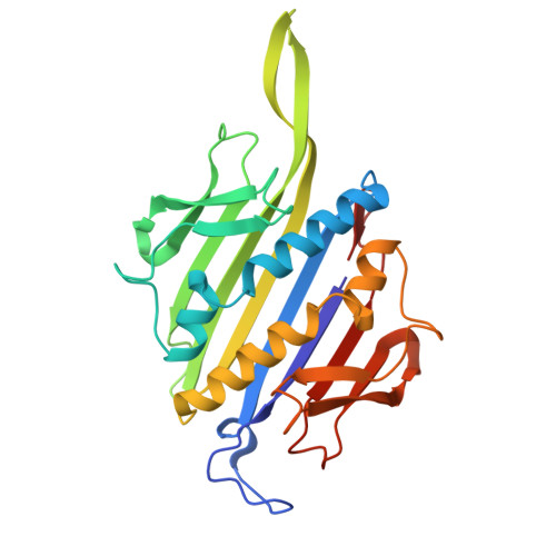
9OH5 - PubMed Abstract:
Virus-like particles (VLPs) are self-assembling nanoparticles derived from viruses with the potential as scaffolds for myriad applications. They are also excellent testbeds for engineering protein superstructures. Engineers often employ techniques such as amino acid substitutions and insertions/deletions. Yet evolution also utilizes circular permutation, a powerful natural strategy that has not been fully explored in engineering self-assembling protein nanoparticles. Here, we demonstrate this technique using the MS2 VLP as a model self-assembling proteinaceous nanoparticle. We constructed a comprehensive circular permutation library of the fused MS2 coat protein dimer construct. The strategy revealed terminal locations, validated via cryo-electron microscopy, that enabled C-terminal peptide tagging and led to a protein encapsulation strategy via covalent bonding - a feature the native coat protein does not permit. Our systematic study demonstrates the power of circular permutation for engineering features as well as quantitatively and systematically exploring VLP structural determinants.
- Interdisciplinary Biological Sciences Program, Northwestern University, Evanston, Illinois 60208, United States.
Organizational Affiliation: