The unique architecture of umbrella toxins permits a two-tiered molecular bet-hedging strategy for interbacterial antagonism.
Zhao, Q., Vlach, J., Park, Y.J., Tan, Y., Bertolli, S.K., Srinivas, P., Liao, P., Fitzpatrick, C.R., Dangl, J.L., Azadi, P., DiMaio, F., Peterson, S.B., Zhang, D., Veesler, D., Mougous, J.D.(2025) Cell
- PubMed: 41338195 Search on PubMed
- DOI: https://doi.org/10.1016/j.cell.2025.10.044
- Primary Citation Related Structures:
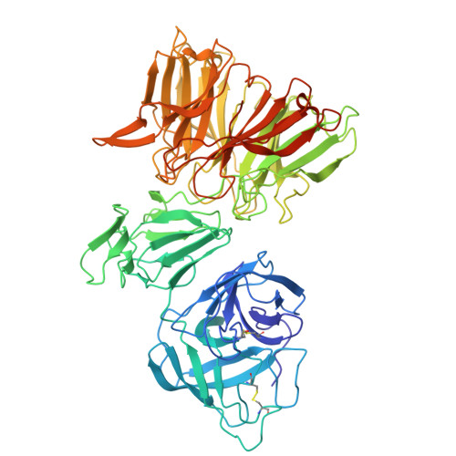
9OEE - PubMed Abstract:
Bacteria exist in competitive and rapidly changing environments in which the nature of future threats cannot be easily predicted. Streptomyces coelicolor produces three antibacterial umbrella particles that harbor distinct polymorphic toxin domains and an overlapping set of six diversified lectins. Here, we show that the exquisite specificity of umbrella particles derives from lectin-mediated species-specific binding to previously undescribed hypervariable surface glycoconjugates. A cryo-electron microscopy (cryo-EM) structure of one such lectin in complex with its oligosaccharide substrate defines the molecular basis for targeting through the coordinated recognition of multiple glycan features. Biochemical and genetic studies of several target species, in conjunction with lectin-swapping experiments, support a model whereby S. coelicolor umbrella toxin diversification at the levels of lectin composition and toxin polymorphism represents a unique, two-tiered bet-hedging strategy. Bioinformatic analyses support this as a means by which the unusual architecture of umbrella toxins offers Streptomyces a generalizable strategy to antagonize an unpredictable array of competitors.
- Department of Microbiology, University of Washington, Seattle, WA, USA.
Organizational Affiliation: