9ODT | pdb_00009odt

The structure of a Bacterial Cyanide Dihydratase from Bacillus safensis PER-URP-08


Experimental Data Snapshot

  • Method: ELECTRON MICROSCOPY
  • Resolution: 2.04 Å
  • Aggregation State: PARTICLE 
  • Reconstruction Method: SINGLE PARTICLE 

wwPDB Validation 3D Report Full Report

Validation slider image for 9ODT

This is version 1.1 of the entry. See complete history

Literature

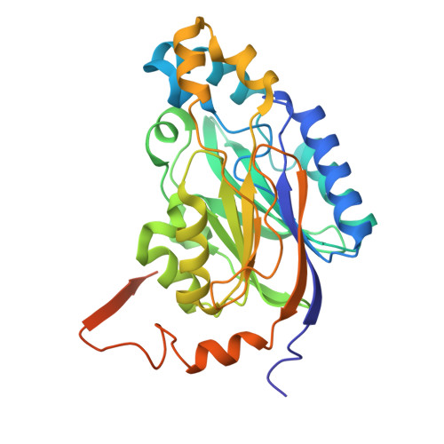
The single-particle cryo-EM structures of a bacterial cyanide dihydratase and a fungal cyanide hydratase.

Justo Arevalo, S.Valle-Riestra Felice, V.Barahona Acuna, M.Ordinola Flores, K.Quinones Aguilar, M.Balan, A.Farah, C.S.

(2026) Structure 34: 599-610.e2

  • DOI: https://doi.org/10.1016/j.str.2026.01.009
  • Primary Citation Related Structures: 
    9ODT, 9OFA

  • PubMed Abstract: 

    Cyanide is widely used in industries due to its affinity for metals, a property that also underlies its toxicity. Industries, therefore, must reduce cyanide concentration before the final disposal of wastewater. Physical, chemical, and biological methods have been developed for this; however, knowledge about the structure of enzymes involved in cyanide degradation remains limited. Structural characterization of these proteins could facilitate the development of enzymes with enhanced bioremediation potential. Here, we present the single-particle cryo-electron microscopy structures of a cyanide dihydratase from Bacillus safensis and a cyanide hydratase from Gloeocercospora sorghi at 2.2 Å and 2.0 Å resolution, respectively. We provide a comprehensive description and comparative analysis alongside all previously determined nitrilase structures. Importantly, our full-length structures reveal new features in the C-terminal as well as specific intermolecular interactions between protomer interfaces and within the helix lumen. Finally, our findings offer insights into the reaction mechanisms of these two enzymes.


  • Organizational Affiliation
    • Departamento de Bioquimica, Instituto de Quimica - Universidade de São Paulo, São Paulo, Brazil; Facultad de Ciencias Biologicas - Universidad Ricardo Palma, Santiago de Surco, Peru. Electronic address: santiago.jus.are@usp.br.

Macromolecule Content 

  • Total Structure Weight: 675.47 kDa 
  • Atom Count: 45,542 
  • Modeled Residue Count: 5,712 
  • Deposited Residue Count: 5,940 
  • Unique protein chains: 1

Macromolecules

Find similar proteins by:|  3D Structure
Entity ID: 1
MoleculeChains  Sequence LengthOrganismDetailsImage
Cyanide dihydratase330Bacillus safensisMutation(s): 0 
Gene Names: cynDEGI07_08135
EC: 3.5.1.49 (PDB Primary Data), 4.2.1.66 (PDB Primary Data)
UniProt
Find proteins for P0DXY3 (Bacillus safensis)
Explore P0DXY3 
Go to UniProtKB:  P0DXY3
Entity Groups
Sequence Clusters30% Identity50% Identity70% Identity90% Identity95% Identity100% Identity
UniProt GroupP0DXY3
Sequence Annotations
Expand
Reference Sequence

Experimental Data & Validation

Experimental Data

  • Method: ELECTRON MICROSCOPY
  • Resolution: 2.04 Å
  • Aggregation State: PARTICLE 
  • Reconstruction Method: SINGLE PARTICLE 
EM Software:
TaskSoftware PackageVersion
RECONSTRUCTIONcryoSPARC4.6.2

Structure Validation

View Full Validation Report


Entry History 

& Funding Information

Deposition Data


Funding OrganizationLocationGrant Number
Sao Paulo Research Foundation (FAPESP)Brazil2022/00943-1
Sao Paulo Research Foundation (FAPESP)Brazil2021/10577-0

Revision History  (Full details and data files)

  • Version 1.0: 2026-02-25
    Type: Initial release
  • Version 1.1: 2026-04-15
    Changes: Data collection, Database references, Source and taxonomy, Structure summary