9O8Z | pdb_00009o8z

CryoEM structure of M. mazei topoisomerase VI-minicircle DNA complex in partially unfolded transducer state


Experimental Data Snapshot

  • Method: ELECTRON MICROSCOPY
  • Resolution: 4.00 Å
  • Aggregation State: PARTICLE 
  • Reconstruction Method: SINGLE PARTICLE 

Starting Models: experimental, other
View more details

wwPDB Validation   3D Report Full Report


This is version 1.1 of the entry. See complete history


Literature

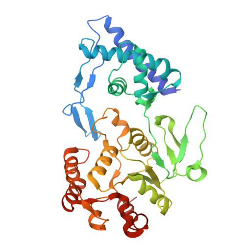
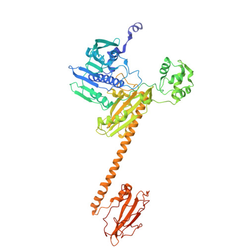
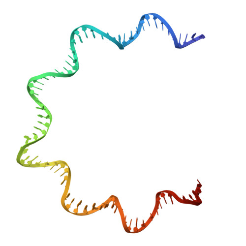
Supercoiled DNA recognition and cleavage control in topoisomerase VI.

Richman, D.E.Wendorff, T.J.Rashid, F.Beck, C.Yan, Q.Johnson, H.R.Eckerty, R.A.Fogg, J.M.Baker, M.L.Zechiedrich, L.Berger, J.M.

(2026) Nat Commun 

  • DOI: https://doi.org/10.1038/s41467-026-69491-0
  • Primary Citation Related Structures: 
    9O0G, 9O7O, 9O8P, 9O8Z, 9O9M

  • PubMed Abstract: 

    Type II topoisomerases modulate DNA supercoiling and resolve chromosome entanglements. Type IIB topoisomerases, exemplified by DNA topoisomerase VI (Top6), are used by plants and archaea to support endoreduplication and cell proliferation, respectively; homologs of Top6 further serve to initiate meiotic recombination in eukaryotes and constitute the nuclease portion of MksBEFG/Wadjet/Gabija bacterial defense systems. To understand how such factors act upon DNA, we determine structures of Top6 bound to supercoiled minicircles in cleaved and uncleaved states using single-particle electron cryo-microscopy. The structures show that Top6 binds a curved 74 bp region of the supercoiled minicircle DNA and that it cuts at a distinct deformability motif, explaining its preference for supercoiled substrates and highlighting the role of DNA plasticity in cleavage site selection. Dynamic protein-DNA interactions and an unanticipated tension sensor help recognize bent DNA and couple ATPase disposition to cleavage state activation. Our observations explain how DNA recognition and cleavage by type II topoisomerases are regulated by interdependent structural changes in DNA and the enzyme.


  • Organizational Affiliation
    • Department of Biophysics & Biophysical Chemistry, Johns Hopkins University School of Medicine, Baltimore, MD, USA.

Macromolecules

Find similar proteins by:  (by identity cutoff)  |  3D Structure
Entity ID: 1
MoleculeChains Sequence LengthOrganismDetailsImage
Type 2 DNA topoisomerase 6 subunit A
A, C
369Methanosarcina mazei Go1Mutation(s): 0 
Gene Names: top6AMM_2418
EC: 5.6.2.2
UniProt
Find proteins for Q8PUB7 (Methanosarcina mazei (strain ATCC BAA-159 / DSM 3647 / Goe1 / Go1 / JCM 11833 / OCM 88))
Explore Q8PUB7 
Go to UniProtKB:  Q8PUB7
Entity Groups  
Sequence Clusters30% Identity50% Identity70% Identity90% Identity95% Identity100% Identity
UniProt GroupQ8PUB7
Sequence Annotations
Expand
  • Reference Sequence
Find similar proteins by:  (by identity cutoff)  |  3D Structure
Entity ID: 2
MoleculeChains Sequence LengthOrganismDetailsImage
Type 2 DNA topoisomerase 6 subunit B
B, D
621Methanosarcina mazei Go1Mutation(s): 0 
Gene Names: top6BMM_2417
EC: 5.6.2.2
UniProt
Find proteins for Q8PUB8 (Methanosarcina mazei (strain ATCC BAA-159 / DSM 3647 / Goe1 / Go1 / JCM 11833 / OCM 88))
Explore Q8PUB8 
Go to UniProtKB:  Q8PUB8
Entity Groups  
Sequence Clusters30% Identity50% Identity70% Identity90% Identity95% Identity100% Identity
UniProt GroupQ8PUB8
Sequence Annotations
Expand
  • Reference Sequence
Find similar nucleic acids by:  (by identity cutoff) 
Entity ID: 3
MoleculeChains LengthOrganismImage
Minicircle DNA (arbitrary model sequence)E [auth F],
F [auth H]
74unidentified
Sequence Annotations
Expand
  • Reference Sequence
Small Molecules
Ligands 3 Unique
IDChains Name / Formula / InChI Key2D Diagram3D Interactions
ANP
Query on ANP

Download Ideal Coordinates CCD File 
H [auth B],
K [auth D]
PHOSPHOAMINOPHOSPHONIC ACID-ADENYLATE ESTER
C10 H17 N6 O12 P3
PVKSNHVPLWYQGJ-KQYNXXCUSA-N
K
Query on K

Download Ideal Coordinates CCD File 
J [auth B],
M [auth D]
POTASSIUM ION
K
NPYPAHLBTDXSSS-UHFFFAOYSA-N
MG
Query on MG

Download Ideal Coordinates CCD File 
G [auth A],
I [auth B],
L [auth D]
MAGNESIUM ION
Mg
JLVVSXFLKOJNIY-UHFFFAOYSA-N
Experimental Data & Validation

Experimental Data

  • Method: ELECTRON MICROSCOPY
  • Resolution: 4.00 Å
  • Aggregation State: PARTICLE 
  • Reconstruction Method: SINGLE PARTICLE 
EM Software:
TaskSoftware PackageVersion
MODEL REFINEMENTPHENIX1.21.1_5286
RECONSTRUCTIONcryoSPARC4.4.1
RECONSTRUCTIONEMReady2.0

Structure Validation

View Full Validation Report


Entry History & Funding Information

Deposition Data


Funding OrganizationLocationGrant Number
National Institutes of Health/National Cancer Institute (NIH/NCI)United States5R35 CA263778
National Institutes of Health/National Institute of General Medical Sciences (NIH/NIGMS)United States1F32 GM128269

Revision History  (Full details and data files)

  • Version 1.0: 2026-02-25
    Type: Initial release
  • Version 1.1: 2026-03-04
    Changes: Data collection, Database references