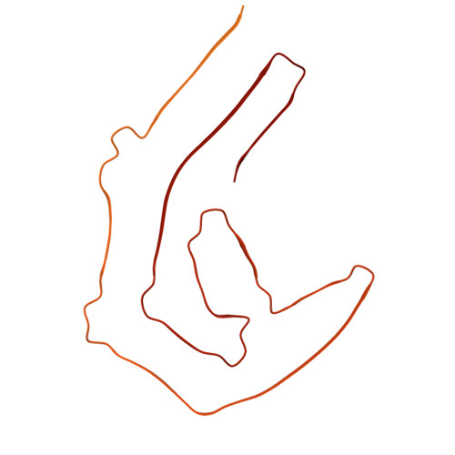
9O8E | pdb_00009o8e

amyloid fibril of recombinant full-length 2N4R tau complexed with unfractionated mouse liver RNA and seeded by Alzheimer's disease tau fibrils


Experimental Data Snapshot

  • Method: ELECTRON MICROSCOPY
  • Resolution: 3.10 Å
  • Aggregation State: FILAMENT 
  • Reconstruction Method: HELICAL 

wwPDB Validation 3D Report Full Report

Validation slider image for 9O8E

This is version 1.1 of the entry. See complete history

Literature

Structural evidence that RNA contributes to polymorphism of tau amyloid fibrils.

Abskharon, R.Jiang, Y.X.Sawaya, M.R.Ge, P.Zhang, J.Boyer, D.R.Dolinsky, J.L.Pi, J.Cascio, D.Guo, F.Eisenberg, D.S.

(2026) iScience 29: 115501-115501

  • DOI: https://doi.org/10.1016/j.isci.2026.115501
  • Primary Citation Related Structures: 
    9O8E, 9O8H

  • PubMed Abstract: 

    RNA colocalizes with tau deposits in Alzheimer's disease (AD) and drives tau aggregation in vitro . Previously, we determined a cryogenic-electron microscopy (cryo-EM) structure of fibrils of full-length tau bound to unfractionated mammalian RNA, revealing a small tau C-terminal core. Here, we present the cryo-EM structure of fibrils of full-length recombinant tau bound to unfractionated mammalian RNA seeded by AD-extracted tau fibrils. This structure reveals an expanded tau C-terminal core resembling AD tau fibrils. RNA sequencing identified 18S ribosomal RNA as the primary fibril-bound species. Next, we determined the cryo-EM structure of fibrils of full-length recombinant tau bound to mammalian 18S ribosomal RNA, revealing a core that consists of the R2 to R4 repeat domains previously seen in pathological tau fibrils. All our recombinant RNA-tau fibrils dissolve upon RNase treatment. Tau fibrils adopt distinct folds in the presence of different RNAs, suggesting RNA is a cofactor capable of shaping tau fibril polymorphism.


  • Organizational Affiliation
    • Department of Chemistry and Biochemistry, Department of Biological Chemistry, UCLA-DOE Institute, Molecular Biology Institute, UCLA, Los Angeles, CA 90095, USA.

Macromolecule Content 

  • Total Structure Weight: 234.96 kDa 
  • Atom Count: 4,165 
  • Modeled Residue Count: 545 
  • Deposited Residue Count: 2,245 
  • Unique protein chains: 1

Macromolecules

Find similar proteins by:|  3D Structure
Entity ID: 1
MoleculeChains  Sequence LengthOrganismDetailsImage
Isoform Tau-F of Microtubule-associated protein tau
A, B, C, D, E
449Homo sapiensMutation(s): 0 
Gene Names: MAPTMAPTLMTBT1TAU
UniProt & NIH Common Fund Data Resources
Find proteins for P10636 (Homo sapiens)
Explore P10636 
Go to UniProtKB:  P10636
PHAROS:  P10636
GTEx:  ENSG00000186868 
Entity Groups
Sequence Clusters30% Identity50% Identity70% Identity90% Identity95% Identity100% Identity
UniProt GroupP10636
Sequence Annotations
Expand
Reference Sequence

Experimental Data & Validation

Experimental Data

  • Method: ELECTRON MICROSCOPY
  • Resolution: 3.10 Å
  • Aggregation State: FILAMENT 
  • Reconstruction Method: HELICAL 
EM Software:
TaskSoftware PackageVersion
MODEL REFINEMENTPHENIX1.21_5207

Structure Validation

View Full Validation Report


Entry History 

& Funding Information

Deposition Data


Funding OrganizationLocationGrant Number
National Institutes of Health/National Institute on Aging (NIH/NIA)United StatesR01 AG070895
National Institutes of Health/National Institute on Aging (NIH/NIA)United StatesRF1 AG065407

Revision History  (Full details and data files)

  • Version 1.0: 2026-03-18
    Type: Initial release
  • Version 1.1: 2026-05-06
    Changes: Data collection, Database references