9O0R | pdb_00009o0r

Crystal structure of wild-type KRAS (GDP-bound) in complex with MRTX849 (adagrasib)


Experimental Data Snapshot

  • Method: X-RAY DIFFRACTION
  • Resolution: 1.81 Å
  • R-Value Free: 
    0.247 (Depositor), 0.281 (DCC) 
  • R-Value Work: 
    0.206 (Depositor) 
  • R-Value Observed: 
    0.209 (Depositor) 

Starting Model: experimental
View more details

wwPDB Validation 3D Report Full Report

Validation slider image for 9O0R

Ligand Structure Quality Assessment 


This is version 1.1 of the entry. See complete history

Literature

Biophysical and structural analysis of KRAS switch-II pocket inhibitors reveals allele-specific binding constraints.

Alexander, P.Chan, A.H.Rabara, D.Swain, M.Larsen, E.K.Dyba, M.Chertov, O.Ashraf, M.Champagne, A.Lin, K.Maciag, A.Gillette, W.K.Nissley, D.V.McCormick, F.Simanshu, D.K.Stephen, A.G.

(2025) J Biological Chem 301: 110331-110331

  • DOI: https://doi.org/10.1016/j.jbc.2025.110331
  • Primary Citation Related Structures: 
    9O0R, 9O0S

  • PubMed Abstract: 

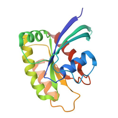
    RAS mutations are observed in 20% of all cancers, with the KRAS isoform highly mutated in colorectal, lung and pancreatic cancers. The last several years have seen the development of clinical compounds that target KRAS G12C mutations, with other compounds under clinical development. In this study, a series of KRAS small-molecule inhibitors were compared for their binding affinity against a panel of KRAS mutant alleles. These inhibitors either covalently target the G12C mutant or reversibly target other mutants by binding in a transient pocket known as the switch-II pocket. Covalent inhibitors bound KRAS-GDP with K D values ranging from 10 -9 -10 -3 M, whereas reversible inhibitors bound in the low nM range. A loss of affinity was observed for KRAS-GppNHp, due in part to rearrangements in switch-II, where the hydrogen bond between G60 and the γ-phosphate needs to break to form the switch-II pocket. Interestingly, these inhibitors had reduced affinity to KRAS Q61R-GppNHp, but not to WT and other mutants. The crystal structure of KRAS Q61R-GppNHp reported here revealed that access to the switch-II pocket was restricted due to R61 forming an additional hydrogen bond with the backbone carbonyl of T35 in switch-I. The restricted access to the switch-II pocket caused a decrease in the association rate of inhibitor binding and resulted in a loss of affinity. These findings across KRAS mutants provide valuable insights into the conformational adaptability of the switch-II pocket and may prove useful in developing the next generation of allele-specific and pan-KRAS small molecule inhibitors.


  • Organizational Affiliation
    • NCI RAS Initiative, Cancer Research Technology Program, Frederick National Laboratory for Cancer Research, Leidos Biomedical Research, Inc., Frederick, MD, USA.

Macromolecule Content 

  • Total Structure Weight: 41.26 kDa 
  • Atom Count: 2,916 
  • Modeled Residue Count: 334 
  • Deposited Residue Count: 340 
  • Unique protein chains: 1

Macromolecules

Find similar proteins by:|  3D Structure
Entity ID: 1
MoleculeChains  Sequence LengthOrganismDetailsImage
GTPase KRas
A, B
170Homo sapiensMutation(s): 0 
Gene Names: KRASKRAS2RASK2
EC: 3.6.5.2
UniProt & NIH Common Fund Data Resources
Find proteins for P01116 (Homo sapiens)
Explore P01116 
Go to UniProtKB:  P01116
PHAROS:  P01116
GTEx:  ENSG00000133703 
Entity Groups
Sequence Clusters30% Identity50% Identity70% Identity90% Identity95% Identity100% Identity
UniProt GroupP01116
Sequence Annotations
Expand
Reference Sequence

Small Molecules

Ligands 4 Unique
IDChains Name / Formula / InChI Key2D Diagram3D Interactions
A1B7W
(Subject of Investigation/LOI)

Query on A1B7W


Download:Ideal Coordinates CCD File
E [auth A],
K [auth B]
Adagrasib
C32 H35 Cl F N7 O2
PEMUGDMSUDYLHU-ZEQRLZLVSA-N
GDP

Query on GDP


Download:Ideal Coordinates CCD File
C [auth A],
I [auth B]
GUANOSINE-5'-DIPHOSPHATE
C10 H15 N5 O11 P2
QGWNDRXFNXRZMB-UUOKFMHZSA-N
ZN

Query on ZN


Download:Ideal Coordinates CCD File
D [auth A]
G [auth A]
H [auth A]
J [auth B]
M [auth B]
D [auth A],
G [auth A],
H [auth A],
J [auth B],
M [auth B],
N [auth B]
ZINC ION
Zn
PTFCDOFLOPIGGS-UHFFFAOYSA-N
ACT

Query on ACT


Download:Ideal Coordinates CCD File
F [auth A],
L [auth B]
ACETATE ION
C2 H3 O2
QTBSBXVTEAMEQO-UHFFFAOYSA-M

Experimental Data & Validation

Experimental Data

  • Method: X-RAY DIFFRACTION
  • Resolution: 1.81 Å
  • R-Value Free:  0.247 (Depositor), 0.281 (DCC) 
  • R-Value Work:  0.206 (Depositor) 
  • R-Value Observed: 0.209 (Depositor) 
Space Group: P 1
Unit Cell:
Length ( Å )Angle ( ˚ )
a = 40.88α = 94.382
b = 42.598β = 98.61
c = 54.483γ = 106.701
Software Package:
Software NamePurpose
PHENIXrefinement
Aimlessdata scaling
XDSdata reduction
PHASERphasing

Structure Validation

View Full Validation Report


Ligand Structure Quality Assessment 


Entry History 

& Funding Information

Deposition Data


Funding OrganizationLocationGrant Number
National Institutes of Health/National Cancer Institute (NIH/NCI)United States75N91019D00024

Revision History  (Full details and data files)

  • Version 1.0: 2025-06-25
    Type: Initial release
  • Version 1.1: 2025-07-16
    Changes: Database references