


Performance of low-voltage cryo-electron microscopes
Choi, H., Hou, C.F.D., Firnberg, E., Wei, H., Petrou, V.I., Kaelber, J.T.To be published.
Experimental Data Snapshot
Starting Model: experimental
View more details
wwPDB Validation 3D Report Full Report

Entity ID: 1 | |||||
|---|---|---|---|---|---|
| Molecule | Chains | Sequence Length | Organism | Details | Image |
| Capsid protein VP1 | 518 | Adeno-associated virus 9 | Mutation(s): 3 Gene Names: cap | 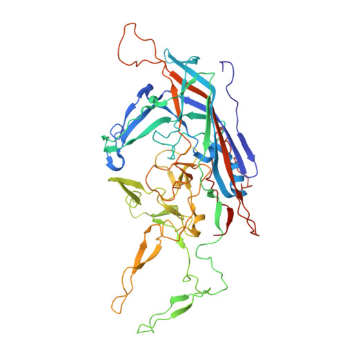 | |
UniProt | |||||
Entity Groups | |||||
| Sequence Clusters | 30% Identity50% Identity70% Identity90% Identity95% Identity100% Identity | ||||
| UniProt Group | Q6JC40 | ||||
Sequence AnnotationsExpand | |||||
Reference Sequence | |||||
| Task | Software Package | Version |
|---|---|---|
| RECONSTRUCTION | cryoSPARC | 4 |
| MODEL REFINEMENT | PHENIX | 1.19.2 |
| Funding Organization | Location | Grant Number |
|---|---|---|
| Not funded | -- |


