9NWW | pdb_00009nww

Single-particle cryo-EM structure of the first variant of mobilized colistin resistance (MCR-1) in its ligand-bound state


Experimental Data Snapshot

  • Method: ELECTRON MICROSCOPY
  • Resolution: 3.58 Å
  • Aggregation State: PARTICLE 
  • Reconstruction Method: SINGLE PARTICLE 

wwPDB Validation   3D Report Full Report


This is version 1.0 of the entry. See complete history


Literature

Mechanistic basis of antimicrobial resistance mediated by the phosphoethanolamine transferase MCR-1.

Zinkle, A.P.Batista, M.B.Herrera, C.M.Erramilli, S.K.Kloss, B.Ashraf, K.U.Nosol, K.Zhang, G.Cater, R.J.Marty, M.T.Kossiakoff, A.A.Trent, M.S.Nygaard, R.Stansfeld, P.J.Mancia, F.

(2025) Nat Commun 16: 10516-10516

  • DOI: https://doi.org/10.1038/s41467-025-65515-3
  • Primary Citation of Related Structures:  
    9NWW

  • PubMed Abstract: 

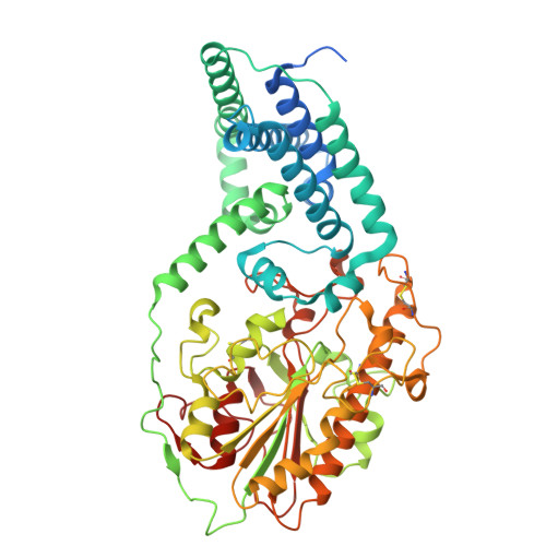
    Polymyxins are used to treat infections caused by multidrug-resistant Gram-negative bacteria. They are cationic peptides that target the negatively charged lipid A component of lipopolysaccharides, disrupting the outer membrane and lysing the cell. Polymyxin resistance is conferred by inner-membrane enzymes, such as phosphoethanolamine transferases, which add positively charged phosphoethanolamine to lipid A. Here, we present the structure of MCR-1, a plasmid-encoded phosphoethanolamine transferase, in its liganded form. The phosphatidylethanolamine donor substrate is bound near the active site in the periplasmic domain, and lipid A is bound over 20 Å away, within the transmembrane region. Integrating structural, biochemical, and drug-resistance data with computational analyses, we propose a two-state model in which the periplasmic domain rotates to bring the active site to lipid A, near the preferential phosphate modification site for MCR-1. This enzymatic mechanism may be generally applicable to other phosphoform transferases with large, globular soluble domains.


  • Organizational Affiliation
    • Department of Physiology and Cellular Biophysics, Columbia University Irving Medical Center, New York, NY, USA.

Macromolecules
Find similar proteins by:  (by identity cutoff)  |  3D Structure
Entity ID: 1
MoleculeChains Sequence LengthOrganismDetailsImage
Probable phosphatidylethanolamine transferase Mcr-1A [auth B]577Escherichia coliMutation(s): 0 
Gene Names: mcr1mcr-1APZ14_31440
EC: 2.7 (PDB Primary Data), 2.7.8.43 (UniProt)
UniProt
Find proteins for A0A0R6L508 (Escherichia coli)
Explore A0A0R6L508 
Go to UniProtKB:  A0A0R6L508
Entity Groups  
Sequence Clusters30% Identity50% Identity70% Identity90% Identity95% Identity100% Identity
UniProt GroupA0A0R6L508
Sequence Annotations
Expand
  • Reference Sequence
Find similar proteins by:  (by identity cutoff)  |  3D Structure
Entity ID: 2
MoleculeChains Sequence LengthOrganismDetailsImage
Fab (MR6) Light (L) ChainB [auth C]214Homo sapiensMutation(s): 0 
Entity Groups  
Sequence Clusters30% Identity50% Identity70% Identity90% Identity95% Identity100% Identity
Sequence Annotations
Expand
  • Reference Sequence
Find similar proteins by:  (by identity cutoff)  |  3D Structure
Entity ID: 3
MoleculeChains Sequence LengthOrganismDetailsImage
Fab (MR6) Heavy (H) ChainC [auth D]235Homo sapiensMutation(s): 0 
Entity Groups  
Sequence Clusters30% Identity50% Identity70% Identity90% Identity95% Identity100% Identity
Sequence Annotations
Expand
  • Reference Sequence
Small Molecules
Ligands 2 Unique
IDChains Name / Formula / InChI Key2D Diagram3D Interactions
KDL (Subject of Investigation/LOI)
Query on KDL

Download Ideal Coordinates CCD File 
D [auth B](2~{R},4~{R},5~{R},6~{R})-6-[(1~{R})-1,2-bis(oxidanyl)ethyl]-2-[(2~{R},4~{R},5~{R},6~{R})-6-[(1~{R})-1,2-bis(oxidanyl)ethyl]-2-carboxy-2-[[(2~{R},3~{S},4~{R},5~{R},6~{R})-5-[[(3~{R})-3-dodecanoyloxytetradecanoyl]amino]-6-[[(2~{R},3~{S},4~{R},5~{R},6~{R})-3-oxidanyl-5-[[(3~{R})-3-oxidanyltetradecanoyl]amino]-4-[(3~{R})-3-oxidanyltetradecanoyl]oxy-6-phosphonooxy-oxan-2-yl]methoxy]-3-phosphonooxy-4-[(3~{R})-3-tetradecanoyloxytetradecanoyl]oxy-oxan-2-yl]methoxy]-5-oxidanyl-oxan-4-yl]oxy-4,5-bis(oxidanyl)oxane-2-carboxylic acid
C110 H202 N2 O39 P2
DIXUKJUHGLIZGU-OIPVZEHTSA-N
PEE (Subject of Investigation/LOI)
Query on PEE

Download Ideal Coordinates CCD File 
E [auth B]1,2-dioleoyl-sn-glycero-3-phosphoethanolamine
C41 H78 N O8 P
MWRBNPKJOOWZPW-NYVOMTAGSA-N
Experimental Data & Validation

Experimental Data

  • Method: ELECTRON MICROSCOPY
  • Resolution: 3.58 Å
  • Aggregation State: PARTICLE 
  • Reconstruction Method: SINGLE PARTICLE 
EM Software:
TaskSoftware PackageVersion
MODEL REFINEMENTPHENIX1.21.2_5419

Structure Validation

View Full Validation Report


Entry History & Funding Information

Deposition Data


Funding OrganizationLocationGrant Number
National Institutes of Health/National Institute of General Medical Sciences (NIH/NIGMS)United StatesGM132120
National Institutes of Health/National Institute Of Allergy and Infectious Diseases (NIH/NIAID)United StatesAI181556
National Institutes of Health/National Institute Of Allergy and Infectious Diseases (NIH/NIAID)United StatesAI74416
National Institutes of Health/National Institute Of Allergy and Infectious Diseases (NIH/NIAID)United StatesAI176776
National Institutes of Health/National Institute Of Allergy and Infectious Diseases (NIH/NIAID)United StatesAI150098

Revision History  (Full details and data files)

  • Version 1.0: 2025-12-17
    Type: Initial release