9NRN | pdb_00009nrn

Lipoprotein Lipase Helical Filament with 11 nm diameter

  • Classification: HYDROLASE
  • Organism(s): Bos taurus
  • Mutation(s): No 

  • Deposited: 2025-03-14 Released: 2025-08-13 
  • Deposition Author(s): Gunn, K.H., Wheless, A., Neher, S.B.
  • Funding Organization(s): National Institutes of Health/National Heart, Lung, and Blood Institute (NIH/NHLBI), National Institutes of Health/National Institute of General Medical Sciences (NIH/NIGMS)

Experimental Data Snapshot

  • Method: ELECTRON MICROSCOPY
  • Resolution: 4.20 Å
  • Aggregation State: FILAMENT 
  • Reconstruction Method: HELICAL 

Starting Model: experimental
View more details

wwPDB Validation 3D Report Full Report

Validation slider image for 9NRN

This is version 1.1 of the entry. See complete history

Literature

Cryogenic electron tomography reveals helical organization of lipoprotein lipase in storage vesicles.

Gunn, K.H.Wheless, A.Calcraft, T.Kreutzberger, M.El-Houshy, K.Egelman, E.H.Rosenthal, P.B.Neher, S.B.

(2025) Sci Adv 11: eadx8711-eadx8711

  • DOI: https://doi.org/10.1126/sciadv.adx8711
  • Primary Citation Related Structures: 
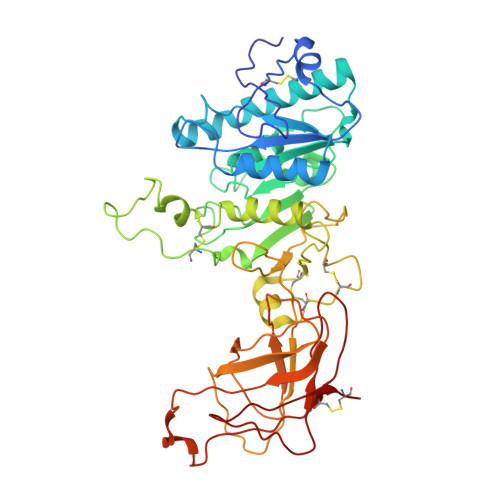
    9NRN

  • PubMed Abstract: 

    Lipoprotein lipase (LPL) is a triglyceride lipase that is contained in intracellular vesicles in an inactive storage form before secretion, but the precise structural details have not yet been resolved. Using cryo-electron tomography (cryo-ET), we observe that LPL exists inside of storage vesicles as a filament with an 11-nanometer diameter and is packed in these vesicles in two distinct patterns. Next, we solved a 4.2-Å resolution cryo-electron microscopy (cryo-EM) structure of this 11-nanometer LPL filament using purified protein. The filament is made of repeating pairs of LPL molecules with occluded active sites, rendering the LPL inactive. The comparison of the in situ subtomogram average and the in vitro cryo-EM structure indicates that the previously uncharacterized physiological storage form of LPL is an inactive filament.


  • Organizational Affiliation
    • Department of Biochemistry and Cell Biology, Stony Brook University, Stony Brook, NY 11790, USA.

Macromolecule Content 

  • Total Structure Weight: 880.23 kDa 
  • Atom Count: 61,884 
  • Modeled Residue Count: 7,812 
  • Deposited Residue Count: 7,812 
  • Unique protein chains: 1

Macromolecules

Find similar proteins by:|  3D Structure
Entity ID: 1
MoleculeChains  Sequence LengthOrganismDetailsImage
Lipoprotein lipase
A, B, C, D, E
A, B, C, D, E, F, G, H, I, J, K, L, M, N, O, P, Q, R
434Bos taurusMutation(s): 0 
EC: 3.1.1.34 (PDB Primary Data), 3.1.1.32 (PDB Primary Data)
UniProt
Find proteins for P11151 (Bos taurus)
Explore P11151 
Go to UniProtKB:  P11151
Entity Groups
Sequence Clusters30% Identity50% Identity70% Identity90% Identity95% Identity100% Identity
UniProt GroupP11151
Sequence Annotations
Expand
Reference Sequence

Experimental Data & Validation

Experimental Data

  • Method: ELECTRON MICROSCOPY
  • Resolution: 4.20 Å
  • Aggregation State: FILAMENT 
  • Reconstruction Method: HELICAL 
EM Software:
TaskSoftware PackageVersion
MODEL REFINEMENTPHENIX1.20.1
RECONSTRUCTIONcryoSPARC

Structure Validation

View Full Validation Report


Entry History 

& Funding Information

Deposition Data


Funding OrganizationLocationGrant Number
National Institutes of Health/National Heart, Lung, and Blood Institute (NIH/NHLBI)United StatesR01 HL125654
National Institutes of Health/National Heart, Lung, and Blood Institute (NIH/NHLBI)United StatesR01 HL163352
National Institutes of Health/National Institute of General Medical Sciences (NIH/NIGMS)United StatesR00 GM146024-04
National Institutes of Health/National Institute of General Medical Sciences (NIH/NIGMS)United StatesGM122510

Revision History  (Full details and data files)

  • Version 1.0: 2025-08-13
    Type: Initial release
  • Version 1.1: 2025-08-20
    Changes: Data collection, Database references