9NRA | pdb_00009nra

The structure of GluA1/A4 LBD-TMD with 4 auxiliary subunits


Experimental Data Snapshot

  • Method: ELECTRON MICROSCOPY
  • Resolution: 4.18 Å
  • Aggregation State: PARTICLE 
  • Reconstruction Method: SINGLE PARTICLE 

wwPDB Validation 3D Report Full Report

Validation slider image for 9NRA

This is version 1.3 of the entry. See complete history

Literature

Gating and noelin clustering of native Ca 2+ -permeable AMPA receptors.

Fang, C.L.Spangler, C.J.Park, J.Sheldon, N.Trussell, L.O.Gouaux, E.

(2025) Nature 645: 526-534

  • DOI: https://doi.org/10.1038/s41586-025-09289-0
  • Primary Citation Related Structures: 
    9NR6, 9NR7, 9NR8, 9NR9, 9NRA

  • PubMed Abstract: 

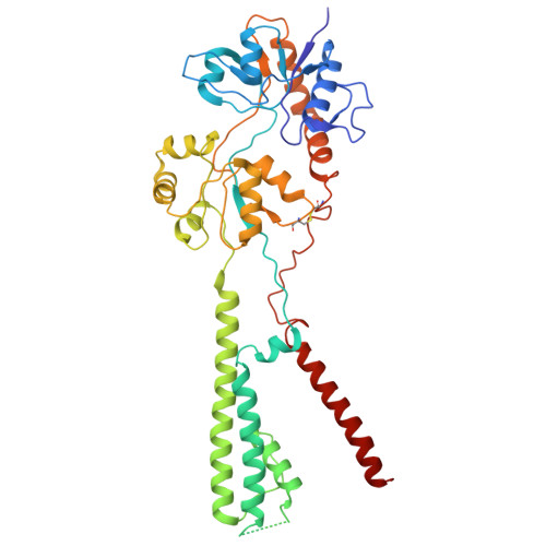
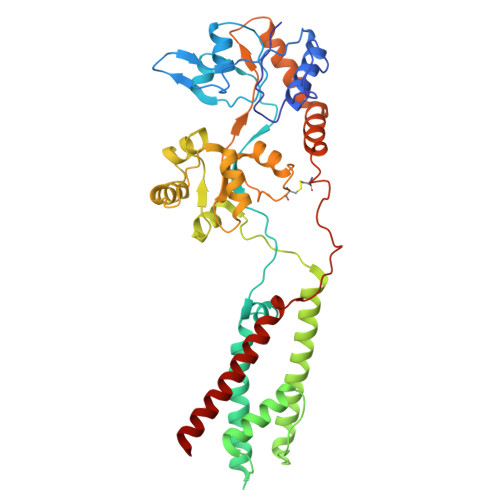
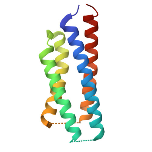
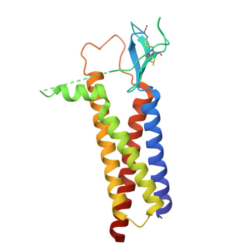
    AMPA-type ionotropic glutamate receptors (AMPARs) are integral to fast excitatory synaptic transmission and play vital roles in synaptic plasticity, motor coordination, learning, and memory 1 . While extensive structural studies have been conducted on recombinant AMPARs and native calcium impermeable (CI)-AMPARs alongside their auxiliary proteins 2-5 , the molecular architecture of native calcium permeable (CP)-AMPARs has remained undefined. To elucidate the subunit composition, physiological architecture, and gating mechanisms of CP-AMPARs, here we present the first visualization of these receptors, immunoaffinity purified from rat cerebella, and resolve their structures using cryo-electron microscopy (cryo-EM). Our results indicate that the predominant assembly consists of GluA1 and GluA4 subunits, with the GluA4 subunit occupying the B and D positions, while auxiliary subunits, including TARPs, are located at the B'/D' positions and CNIHs or TARPs at the A'/C' positions. Furthermore, we resolved the structure of the Noelin 1-GluA1/A4 complex, wherein Noelin 1 (Noe 1) specifically binds to the GluA4 subunit at the B and D positions. Notably, Noe 1 stabilizes the amino-terminal domain (ATD) layer without affecting receptor gating properties. Noe 1 contributes to AMPAR function by forming dimeric-AMPAR assemblies that likely engage in extracellular networks, clustering receptors within synaptic environments and modulating receptor responsiveness to synaptic inputs.


  • Organizational Affiliation
    • Vollum Institute, Oregon Health & Science University, Portland, OR, USA.

Macromolecule Content 

  • Total Structure Weight: 256.9 kDa 
  • Atom Count: 15,598 
  • Modeled Residue Count: 2,195 
  • Deposited Residue Count: 2,344 
  • Unique protein chains: 4

Macromolecules

Find similar proteins by:|  3D Structure
Entity ID: 1
MoleculeChains  Sequence LengthOrganismDetailsImage
Glutamate receptor 1
A, C
427Rattus norvegicusMutation(s): 0 
UniProt
Find proteins for P19490 (Rattus norvegicus)
Explore P19490 
Go to UniProtKB:  P19490
Entity Groups
Sequence Clusters30% Identity50% Identity70% Identity90% Identity95% Identity100% Identity
UniProt GroupP19490
Sequence Annotations
Expand
Reference Sequence
Find similar proteins by:|  3D Structure
Entity ID: 2
MoleculeChains  Sequence LengthOrganismDetailsImage
Isoform 2 of Glutamate receptor 4
B, D
424Rattus norvegicusMutation(s): 0 
UniProt
Find proteins for P19493 (Rattus norvegicus)
Explore P19493 
Go to UniProtKB:  P19493
Entity Groups
Sequence Clusters30% Identity50% Identity70% Identity90% Identity95% Identity100% Identity
UniProt GroupP19493
Sequence Annotations
Expand
Reference Sequence
Find similar proteins by:|  3D Structure
Entity ID: 3
MoleculeChains  Sequence LengthOrganismDetailsImage
Auxiliary protein at A'/C'
E, G
117Rattus norvegicusMutation(s): 0 
Find similar proteins by:|  3D Structure
Entity ID: 4
MoleculeChains  Sequence LengthOrganismDetailsImage
Voltage-dependent calcium channel gamma-2 subunit
F, H
204Rattus norvegicusMutation(s): 0 
UniProt
Find proteins for Q71RJ2 (Rattus norvegicus)
Explore Q71RJ2 
Go to UniProtKB:  Q71RJ2
Entity Groups
Sequence Clusters30% Identity50% Identity70% Identity90% Identity95% Identity100% Identity
UniProt GroupQ71RJ2
Sequence Annotations
Expand
Reference Sequence

Experimental Data & Validation

Experimental Data

  • Method: ELECTRON MICROSCOPY
  • Resolution: 4.18 Å
  • Aggregation State: PARTICLE 
  • Reconstruction Method: SINGLE PARTICLE 
EM Software:
TaskSoftware PackageVersion
MODEL REFINEMENTPHENIX1.20.1_4487:

Structure Validation

View Full Validation Report


Entry History 

& Funding Information

Deposition Data


Funding OrganizationLocationGrant Number
Howard Hughes Medical Institute (HHMI)United States--

Revision History  (Full details and data files)

  • Version 1.0: 2025-07-02
    Type: Initial release
  • Version 1.1: 2025-07-09
    Changes: Data collection, Database references
  • Version 1.2: 2025-09-17
    Changes: Data collection, Database references
  • Version 1.3: 2025-11-26
    Changes: Data collection, Database references, Structure summary