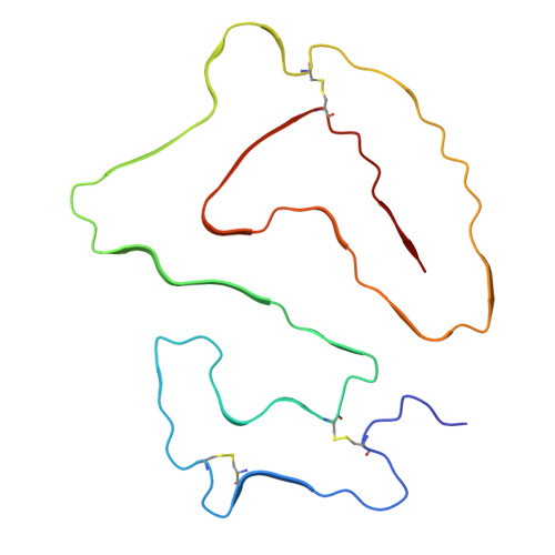
9NON | pdb_00009non

Cryo-EM structure of amyloid fibril isolated from the kidney of a variant Alect2-I40V amyloidosis patient

  • Classification: PROTEIN FIBRIL
  • Organism(s): Homo sapiens
  • Mutation(s): Yes 

  • Deposited: 2025-03-10 Released: 2026-02-11 
  • Deposition Author(s): Nguyen, B., Saelices, L.
  • Funding Organization(s): American Heart Association, National Institutes of Health/National Heart, Lung, and Blood Institute (NIH/NHLBI), Welch Foundation

Experimental Data Snapshot

  • Method: ELECTRON MICROSCOPY
  • Resolution: 2.40 Å
  • Aggregation State: FILAMENT 
  • Reconstruction Method: HELICAL 

wwPDB Validation 3D Report Full Report

Validation slider image for 9NON

This is version 1.1 of the entry. See complete history

Literature

Structural polymorphism of ex-vivo ALECT2 amyloid fibrils revealed by cryo-EM.

Afrin, S.Nguyen, B.A.Singh, V.Singh, P.Bassett, P.Pekala, M.Evers, B.Lopez, C.Ahmed, Y.Li, L.Kallem, R.R.Lemoff, A.Argyropoulos, C.Kluve-Beckerman, B.Saelices, L.

(2026) Nat Commun 

  • DOI: https://doi.org/10.1038/s41467-026-71223-3
  • Primary Citation Related Structures: 
    9NON

  • PubMed Abstract: 

    ALECT2 amyloidosis is a rare systemic disease characterized by the pathological deposition of leukocyte cell-derived chemotaxin-2 (LECT2) as amyloid fibrils, primarily affecting the kidneys and liver. The molecular mechanisms underlying LECT2 aggregation remain poorly defined, hindering diagnostic and therapeutic development. Here, we present cryo-electron microscopy structures of ex-vivo ALECT2 fibrils extracted from a patient's kidney. We identified three fibril polymorphs: a predominant single-protofilament morphology and two minor double-protofilament morphologies. The dominant single-protofilament morphology comprises the full-length 133-residue LECT2 protein and retains all three native disulfide bonds. Low-resolution reconstructions of double-protofilament morphologies suggest they adopt a similar fold to the single protofilament morphology, but form paired assemblies with different inter-filament interfaces. Mass spectrometry also reveals acetylation within the fibrils. These findings offer critical insights into the structural basis of ALECT2 amyloid formation and identify molecular features that could inform future diagnostic and therapeutic approaches.


  • Organizational Affiliation
    • Center for Alzheimer's and Neurodegenerative Diseases, Department of Biophysics, Peter O'Donnell Jr Brain Institute, University of Texas Southwestern Medical Center (UTSW), Dallas, TX, USA.

Macromolecule Content 

  • Total Structure Weight: 72.97 kDa 
  • Atom Count: 5,105 
  • Modeled Residue Count: 665 
  • Deposited Residue Count: 665 
  • Unique protein chains: 1

Macromolecules

Find similar proteins by:|  3D Structure
Entity ID: 1
MoleculeChains  Sequence LengthOrganismDetailsImage
Leukocyte cell-derived chemotaxin-2A [auth B],
B [auth C],
C [auth A],
D,
E
133Homo sapiensMutation(s): 1 
UniProt & NIH Common Fund Data Resources
Find proteins for O14960 (Homo sapiens)
Explore O14960 
Go to UniProtKB:  O14960
PHAROS:  O14960
GTEx:  ENSG00000145826 
Entity Groups
Sequence Clusters30% Identity50% Identity70% Identity90% Identity95% Identity100% Identity
UniProt GroupO14960
Sequence Annotations
Expand
Reference Sequence

Experimental Data & Validation

Experimental Data

  • Method: ELECTRON MICROSCOPY
  • Resolution: 2.40 Å
  • Aggregation State: FILAMENT 
  • Reconstruction Method: HELICAL 
EM Software:
TaskSoftware PackageVersion
RECONSTRUCTIONRELION4.0
MODEL REFINEMENTPHENIX1.20.1_4487

Structure Validation

View Full Validation Report


Entry History 

& Funding Information

Deposition Data


Funding OrganizationLocationGrant Number
American Heart AssociationUnited States847236
National Institutes of Health/National Heart, Lung, and Blood Institute (NIH/NHLBI)United StatesDP2-HL163810
Welch FoundationUnited StatesI-2121-20220331

Revision History  (Full details and data files)

  • Version 1.0: 2026-02-11
    Type: Initial release
  • Version 1.1: 2026-04-29
    Changes: Data collection, Database references