9NNU | pdb_00009nnu

Crystal Structure of Ebola Envelope glycoprotein GP in complex with compound LD4-189ZbR


Experimental Data Snapshot

  • Method: X-RAY DIFFRACTION
  • Resolution: 2.59 Å
  • R-Value Free: 
    0.244 (Depositor), 0.237 (DCC) 
  • R-Value Work: 
    0.220 (Depositor), 0.219 (DCC) 
  • R-Value Observed: 
    0.222 (Depositor) 

Starting Model: experimental
View more details

wwPDB Validation 3D Report Full Report

Validation slider image for 9NNU

Ligand Structure Quality Assessment 


This is version 1.0 of the entry. See complete history

Literature

SuFEx-enabled high-throughput medicinal chemistry for developing potent tamoxifen analogs as Ebola virus entry inhibitors.

Dada, L.Nagai, E.Agrawal, S.Wirchnianski, A.S.Wilson, I.A.Chandran, K.Kitamura, S.

(2025) Front Immunol 16: 1533037-1533037

  • DOI: https://doi.org/10.3389/fimmu.2025.1533037
  • Primary Citation Related Structures: 
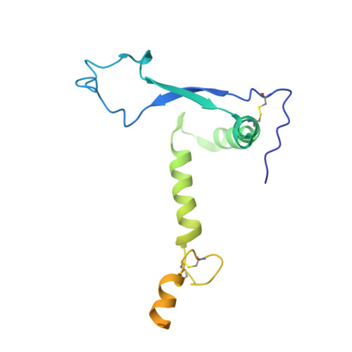
    9NNU

  • PubMed Abstract: 

    Ebola virus (EBOV) causes severe hemorrhagic fever with a high mortality rate in humans. In acute infection, an abnormal immune response results in excessive inflammatory cytokines and uncontrolled systemic inflammation that can result in organ damage and multi-organ failure. While vaccines and monoclonal antibody therapies are available, there is an urgent need for effective small-molecule antivirals against EBOV. Here, we report on the optimization of tamoxifen, an EBOV-glycoprotein (GP) binder that inhibits viral entry, using our Sulfur-Fluoride Exchange (SuFEx) click chemistry-based high-throughput medicinal chemistry (HTMC) strategy. Using a "Direct-to-Biology" approach, we generated a focused library of 2,496 tamoxifen analogs overnight and screened them in a cell-based pseudo-EBOV infection assay. The HTMC workflow enabled the development of a potent EBOV entry inhibitor with submicromolar EC 50 cellular antiviral activity and more than 50-fold improvement in binding affinity against EBOV-GP compared to the parent compound. Our findings underscore the use of SuFEx-enabled HTMC for rapidly generating and assessing potential therapeutic candidates against viral and immune-mediated diseases in a cell-based assay.


  • Organizational Affiliation
    • Department of Biochemistry, Albert Einstein College of Medicine, Bronx, NY, United States.

Macromolecule Content 

  • Total Structure Weight: 72.12 kDa 
  • Atom Count: 3,006 
  • Modeled Residue Count: 366 
  • Deposited Residue Count: 638 
  • Unique protein chains: 2

Macromolecules

Find similar proteins by:|  3D Structure
Entity ID: 1
MoleculeChains  Sequence LengthOrganismDetailsImage
Envelope glycoprotein470Ebola virus - Mayinga, Zaire, 1976Mutation(s): 0 
Gene Names: GP
UniProt
Find proteins for Q05320 (Zaire ebolavirus (strain Mayinga-76))
Explore Q05320 
Go to UniProtKB:  Q05320
Entity Groups
Sequence Clusters30% Identity50% Identity70% Identity90% Identity95% Identity100% Identity
UniProt GroupQ05320
Glycosylation
Glycosylation Sites: 4
Sequence Annotations
Expand
Reference Sequence
Find similar proteins by:|  3D Structure
Entity ID: 2
MoleculeChains  Sequence LengthOrganismDetailsImage
GP2168Ebola virus - Mayinga, Zaire, 1976Mutation(s): 0 
Gene Names: GP
UniProt
Find proteins for Q05320 (Zaire ebolavirus (strain Mayinga-76))
Explore Q05320 
Go to UniProtKB:  Q05320
Entity Groups
Sequence Clusters30% Identity50% Identity70% Identity90% Identity95% Identity100% Identity
UniProt GroupQ05320
Glycosylation
Glycosylation Sites: 1
Sequence Annotations
Expand
Reference Sequence

Oligosaccharides

Help  
Entity ID: 3
MoleculeChains Length2D Diagram GlycosylationD Interactions
alpha-D-mannopyranose-(1-6)-beta-D-mannopyranose-(1-4)-2-acetamido-2-deoxy-beta-D-glucopyranose-(1-4)-2-acetamido-2-deoxy-beta-D-glucopyranose
C
4N-Glycosylation
Glycosylation Resources
GlyTouCan: G22573RC
GlyCosmos: G22573RC
GlyGen: G22573RC

Experimental Data & Validation

Experimental Data

  • Method: X-RAY DIFFRACTION
  • Resolution: 2.59 Å
  • R-Value Free:  0.244 (Depositor), 0.237 (DCC) 
  • R-Value Work:  0.220 (Depositor), 0.219 (DCC) 
  • R-Value Observed: 0.222 (Depositor) 
Space Group: H 3 2
Unit Cell:
Length ( Å )Angle ( ˚ )
a = 114.222α = 90
b = 114.222β = 90
c = 306.37γ = 120
Software Package:
Software NamePurpose
PHENIXrefinement
HKL-2000data reduction
HKL-2000data scaling
PHASERphasing

Structure Validation

View Full Validation Report


Ligand Structure Quality Assessment 


Entry History 

& Funding Information

Deposition Data


Funding OrganizationLocationGrant Number
National Institutes of Health/National Institute Of Allergy and Infectious Diseases (NIH/NIAID)United States--

Revision History  (Full details and data files)

  • Version 1.0: 2025-07-30
    Type: Initial release