9NLO | pdb_00009nlo

Escherichia coli Signal Peptidase I Delta 2-76 P84A in complex with lipopeptide inhibitor


Experimental Data Snapshot

  • Method: X-RAY DIFFRACTION
  • Resolution: 2.32 Å
  • R-Value Free: 
    0.234 (Depositor), 0.234 (DCC) 
  • R-Value Work: 
    0.209 (Depositor), 0.213 (DCC) 

Starting Model: experimental
View more details

wwPDB Validation 3D Report Full Report

Validation slider image for 9NLO

Ligand Structure Quality Assessment 


This is version 1.1 of the entry. See complete history

Literature

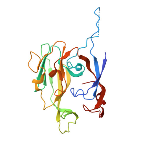
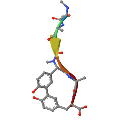
Crystal structure of Escherichia coli type I signal peptidase P84A in complex with lipopeptide antibiotic arylomycin A 2 .

Luo, C.Paetzel, M.

(2025) J Struct Biol 217: 108260-108260

  • DOI: https://doi.org/10.1016/j.jsb.2025.108260
  • Primary Citation Related Structures: 
    9NLO

  • PubMed Abstract: 

    Type I signal peptidase (SPase I) is an essential membrane-bound enzyme that removes amino-terminal signal peptides from secretory proteins. Owing to its critical role in bacterial viability and its periplasmic accessibility, SPase I has emerged as an attractive target for antibiotic development. Arylomycins, a class of macrocyclic lipohexapeptide natural products, inhibit SPase I by binding to its active site. Previous studies have identified a key resistance determinant-a proline residue at the base of the substrate-binding groove (Pro84 inEscherichia coliSPase I)-which reduces arylomycin affinity. Here, we present the crystal structure of theE. coliSPase I P84A mutant in complex with arylomycin A 2 , revealing that the introduced alanine enables an additional hydrogen bond between the enzyme backbone and the arylomycin N-terminal carbonyl, thus enhancing the affinity for arylomycins. Furthermore, a newly developed preprotein-binding assay utilizing a non-cleavable version of ProOmpA Nuclease A demonstrates that substituting SPase I Pro84 with serine or leucine disrupts substrate recognition, underscoring the delicate balance between inhibitor resistance and substrate processing. These findings reveal that residue Pro84 participates in the interaction between preprotein signal peptides and the E. coli SPase I substrate-binding groove, offering a foundation for designing next-generation arylomycin analogs with improved antibacterial potency.


  • Organizational Affiliation
    • Department of Molecular Biology and Biochemistry, Simon Fraser University, South Science Building 8888 University Drive, Burnaby, British Columbia V5A 1S6 Canada.

Macromolecule Content 

  • Total Structure Weight: 57.7 kDa 
  • Atom Count: 3,726 
  • Modeled Residue Count: 467 
  • Deposited Residue Count: 510 
  • Unique protein chains: 2

Macromolecules

Find similar proteins by:|  3D Structure
Entity ID: 1
MoleculeChains  Sequence LengthOrganismDetailsImage
Signal peptidase I
A, B
249Escherichia coli K-12Mutation(s): 1 
Gene Names: lepBb2568JW2552
EC: 3.4.21.89
UniProt
Find proteins for P00803 (Escherichia coli (strain K12))
Explore P00803 
Go to UniProtKB:  P00803
Entity Groups
Sequence Clusters30% Identity50% Identity70% Identity90% Identity95% Identity100% Identity
UniProt GroupP00803
Sequence Annotations
Expand
Reference Sequence
Find similar proteins by:  Sequence   |   3D Structure  
Entity ID: 2
MoleculeChains  Sequence LengthOrganismDetailsImage
ARYLOMYCIN A2
C, D
6synthetic constructMutation(s): 0 

Biologically Interesting Molecules (External Reference) 

1 Unique

Experimental Data & Validation

Experimental Data

  • Method: X-RAY DIFFRACTION
  • Resolution: 2.32 Å
  • R-Value Free:  0.234 (Depositor), 0.234 (DCC) 
  • R-Value Work:  0.209 (Depositor), 0.213 (DCC) 
Space Group: P 43 21 2
Unit Cell:
Length ( Å )Angle ( ˚ )
a = 71.8α = 90
b = 71.8β = 90
c = 263.31γ = 90
Software Package:
Software NamePurpose
REFMACrefinement
MOSFLMdata reduction
SCALAdata scaling
PHASERphasing

Structure Validation

View Full Validation Report


Ligand Structure Quality Assessment 


Entry History 

& Funding Information

Deposition Data


Funding OrganizationLocationGrant Number
Natural Sciences and Engineering Research Council (NSERC, Canada)Canada--

Revision History  (Full details and data files)

  • Version 1.0: 2025-11-19
    Type: Initial release
  • Version 1.1: 2025-11-26
    Changes: Database references