9NE0 | pdb_00009ne0

The flexible portion of Cryo-EM structure of Herpesvirus Helicase-Primase complex prepared with forked DNA, ATP-gamma-S and Pritelivir


Experimental Data Snapshot

  • Method: ELECTRON MICROSCOPY
  • Resolution: 3.40 Å
  • Aggregation State: PARTICLE 
  • Reconstruction Method: SINGLE PARTICLE 

wwPDB Validation   3D Report Full Report


This is version 1.1 of the entry. See complete history


Literature

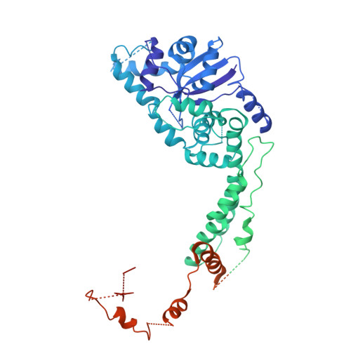
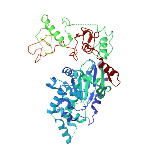
Structural and mechanistic insights into herpesvirus helicase-primase and its therapeutic inhibitors.

Yao, Q.Mercier, A.Nayak, A.May, L.Ho, P.Y.Lewis-Ballester, A.Nair, V.Sapre, A.Aeschbacher, T.Mukherjee, J.Richards, C.Mateo, R.Cho, A.Lansdon, E.Yu, X.

(2025) Nat Microbiol 10: 3191-3201

  • DOI: https://doi.org/10.1038/s41564-025-02168-4
  • Primary Citation of Related Structures:  
    9NDA, 9NDQ, 9NDT, 9NDZ, 9NE0, 9NEB, 9NEE, 9NEL

  • PubMed Abstract: 

    The herpes simplex virus (HSV) helicase-primase (HP) complex is a promising anti-herpes therapeutic target. However, progress in developing highly effective small-molecule HP inhibitors (HPIs) for the treatment of genital herpes has been hindered by the lack of structural information on the HP complex and the incomplete understanding of the mechanism of action of HPIs. Here we present the cryogenic electron microscopy structure of the HSV-1 HP apo-complex (3.8 Å), along with structures bound to pritelivir (3.2 Å) and amenamevir (3.2 Å)-two clinically active, chemically distinct HPIs. The potency of both inhibitors against HSV variants bearing mutations within the HPI binding pocket supports the high-resolution mapping of key molecular interactions while revealing residues that govern their antiviral spectrum against alphaherpesviruses. Our results provide important insight into the unique architecture of the HP complex and the mechanism of inhibition of HPIs, paving the way for the development of next-generation antivirals to treat herpesvirus infections.


  • Organizational Affiliation
    • Gilead Sciences Inc., Foster City, CA, USA. qing.yao1@gilead.com.

Macromolecules
Find similar proteins by:  (by identity cutoff)  |  3D Structure
Entity ID: 1
MoleculeChains Sequence LengthOrganismDetailsImage
DNA primase1,024OrthoherpesviridaeMutation(s): 0 
Gene Names: UL52
EC: 2.7.7
UniProt
Find proteins for P10236 (Human herpesvirus 1 (strain 17))
Explore P10236 
Go to UniProtKB:  P10236
Entity Groups  
Sequence Clusters30% Identity50% Identity70% Identity90% Identity95% Identity100% Identity
UniProt GroupP10236
Sequence Annotations
Expand
  • Reference Sequence
Find similar proteins by:  (by identity cutoff)  |  3D Structure
Entity ID: 2
MoleculeChains Sequence LengthOrganismDetailsImage
DNA replication helicase847OrthoherpesviridaeMutation(s): 0 
Gene Names: HELIUL5
EC: 3.6.4
UniProt
Find proteins for P10189 (Human herpesvirus 1 (strain 17))
Explore P10189 
Go to UniProtKB:  P10189
Entity Groups  
Sequence Clusters30% Identity50% Identity70% Identity90% Identity95% Identity100% Identity
UniProt GroupP10189
Sequence Annotations
Expand
  • Reference Sequence
Small Molecules
Experimental Data & Validation

Experimental Data

  • Method: ELECTRON MICROSCOPY
  • Resolution: 3.40 Å
  • Aggregation State: PARTICLE 
  • Reconstruction Method: SINGLE PARTICLE 
EM Software:
TaskSoftware PackageVersion
MODEL REFINEMENTPHENIX
RECONSTRUCTIONRELION5.0

Structure Validation

View Full Validation Report


Entry History & Funding Information

Deposition Data

  • Released Date: 2025-08-13 
  • Deposition Author(s): Yao, Q., Yu, X.

Funding OrganizationLocationGrant Number
Not funded--

Revision History  (Full details and data files)

  • Version 1.0: 2025-08-13
    Type: Initial release
  • Version 1.1: 2026-02-25
    Changes: Data collection, Database references