Predicting the conformational flexibility of antibody and T cell receptor complementarity-determining regions.
Spoendlin, F.C., Fernandez-Quintero, M.L., Raghavan, S.S.R., Turner, H.L., Gharpure, A., Loeffler, J.R., Wong, W.K., Bujotzek, A., Georges, G., Ward, A.B., Deane, C.M.(2025) Nat Mach Intell 7: 1755-1767
- PubMed: 41143207 Search on PubMedSearch on PubMed Central
- DOI: https://doi.org/10.1038/s42256-025-01131-6
- Primary Citation Related Structures:
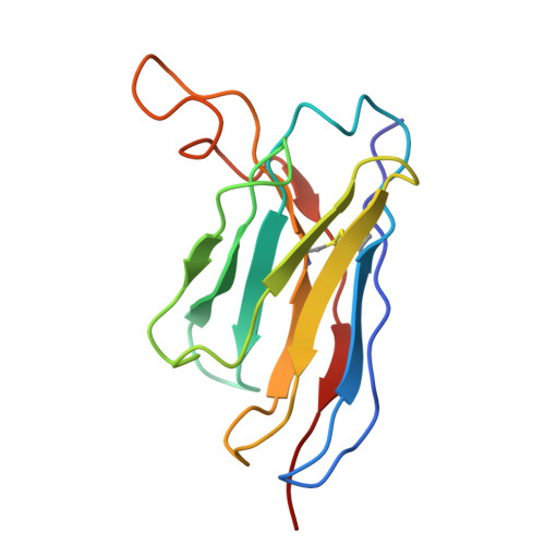
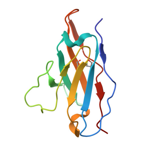
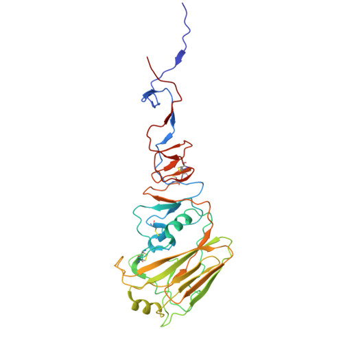
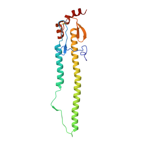
9N5Y, 9N5Z - PubMed Abstract:
Many proteins are highly flexible and their ability to adapt their shape can be fundamental to their functional properties. For example, the flexibility of antibody complementarity-determining region (CDR) loops influences binding affinity and specificity, making it a key factor in understanding and designing antigen interactions. With methods such as AlphaFold, it is possible to computationally predict a single, static protein structure with high accuracy. However, the reliable prediction of structural flexibility has not yet been achieved. A major factor limiting such predictions is the scarcity of suitable training data. Here we focus on predicting the structural flexibility of functionally important antibody and T cell receptor CDR3 loops. To this end, we constructed ALL-conformations by extracting CDR3s and CDR3-like loop motifs from all structures deposited in the Protein Data Bank. This dataset comprises 1.2 million loop structures representing more than 100,000 unique sequences and captures all experimentally observed conformations of these motifs. Using this dataset, we develop ITsFlexible, a deep learning tool with graph neural network architecture. We trained the model to binary classify CDR loops as 'rigid' or 'flexible' from inputs of antibody structures. ITsFlexible outperforms all alternative approaches on our crystal structure datasets and successfully generalizes to molecular dynamics simulations. We also used ITsFlexible to predict the flexibility of three CDRH3 loops with no solved structures and experimentally determined their conformations using cryogenic electron microscopy.
- Department of Statistics, University of Oxford, Oxford, UK.
Organizational Affiliation: