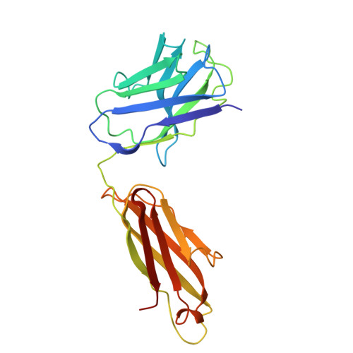
9N5H | pdb_00009n5h

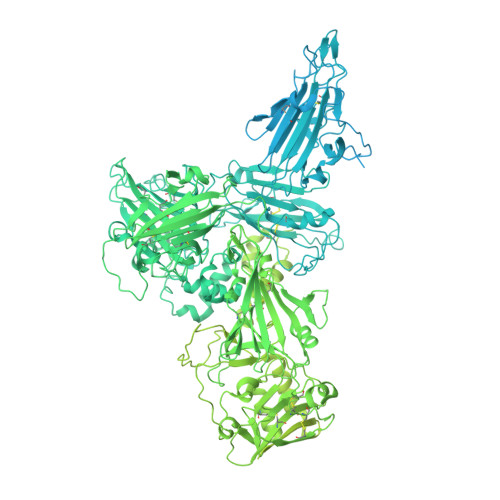
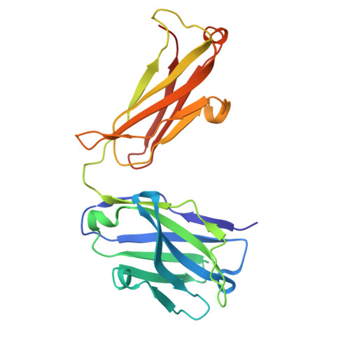
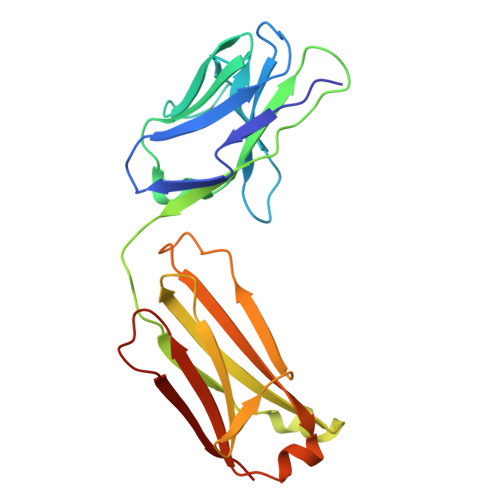
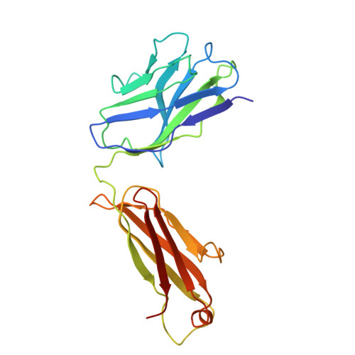
Endogenous Pfs230D1-6 in complex with RUPA-97, LMIV230-01, and 2A2 Fab domains


Experimental Data Snapshot

  • Method: ELECTRON MICROSCOPY
  • Resolution: 3.60 Å
  • Aggregation State: PARTICLE 
  • Reconstruction Method: SINGLE PARTICLE 

Starting Models: in silico, experimental
View more details

wwPDB Validation 3D Report Full Report

Validation slider image for 9N5H

This is version 1.1 of the entry. See complete history. 

Literature

Structure of endogenous Pfs230:Pfs48/45 in complex with potent malaria transmission-blocking antibodies.

Bekkering, E.T., Yoo, R., Hailemariam, S., Heide, F., Ivanochko, D., Jackman, M., Proellochs, N.I., Stoter, R., van Gemert, G.J., Maeda, A., Yuguchi, T., Wanders, O.T., van Daalen, R.C., Inklaar, M.R., Andrade, C.M., Jansen, P.W.T.C., Vermeulen, M., Bousema, T., Takashima, E., Rubinstein, J.L., Kooij, T.W.A., Jore, M.M., Julien, J.P.

(2025) bioRxiv 

  • DOI: https://doi.org/10.1101/2025.02.14.638310
  • Primary Citation Related Structures: 
    9N5H, 9N5K, 9N5O

  • PubMed Abstract: 

    The Pfs230:Pfs48/45 complex forms the basis for leading malaria transmission-blocking vaccine candidates, yet little is known about its molecular assembly. Here, we used cryogenic electron microscopy to elucidate the structure of the endogenous Pfs230:Pfs48/45 complex bound to six potent transmission-blocking antibodies. Pfs230 consists of multiple domain clusters rigidified by interactions mediated through insertion domains. Membrane-anchored Pfs48/45 forms a disc-like structure and interacts with a short C-terminal peptide on Pfs230 that is critical for Pfs230 membrane-retention in vivo . Interestingly, membrane retention through this interaction is not essential for transmission to mosquitoes, suggesting that complex disruption is not a mode of action for transmission-blocking antibodies. Analyses of Pfs48/45- and Pfs230-targeted antibodies identify conserved epitopes on the Pfs230:Pfs48/45 complex and provides a structural paradigm for complement-dependent activity of Pfs230-targeting antibodies. Altogether, the antibody-bound Pfs230:Pfs48/45 structure presented improves our molecular understanding of this biological complex, informing the development of next-generation Plasmodium falciparum transmission-blocking interventions.


  • Organizational Affiliation: 
    • Department of Medical Microbiology, Radboud University Medical Center, The Netherlands.

Macromolecule Content 

  • Total Structure Weight: 505.1 kDa 
  • Atom Count: 19,120 
  • Modeled Residue Count: 2,430 
  • Deposited Residue Count: 4,447 
  • Unique protein chains: 7

Macromolecules

Find similar proteins by:|  3D Structure
Entity ID: 1
MoleculeChains  Sequence LengthOrganismDetailsImage
Gametocyte surface protein P2303,135Plasmodium falciparum 3D7Mutation(s): 0 
UniProt
Find proteins for P68874 (Plasmodium falciparum (isolate 3D7))
Explore P68874 
Go to UniProtKB:  P68874
Entity Groups
Sequence Clusters30% Identity50% Identity70% Identity90% Identity95% Identity100% Identity
UniProt GroupP68874
Sequence Annotations
Expand
Reference Sequence
Find similar proteins by:|  3D Structure
Entity ID: 2
MoleculeChains  Sequence LengthOrganismDetailsImage
2A2 Heavy Chain223Mus musculusMutation(s): 0 
Entity Groups
Sequence Clusters30% Identity50% Identity70% Identity90% Identity95% Identity100% Identity
Sequence Annotations
Expand
Reference Sequence
Find similar proteins by:|  3D Structure
Entity ID: 3
MoleculeChains  Sequence LengthOrganismDetailsImage
2A2 Kappa Chain214Mus musculusMutation(s): 0 
Entity Groups
Sequence Clusters30% Identity50% Identity70% Identity90% Identity95% Identity100% Identity
Sequence Annotations
Expand
Reference Sequence
Find similar proteins by:|  3D Structure
Entity ID: 4
MoleculeChains  Sequence LengthOrganismDetailsImage
LMIV230-01 Heavy Chain221Homo sapiensMutation(s): 0 
Entity Groups
Sequence Clusters30% Identity50% Identity70% Identity90% Identity95% Identity100% Identity
Sequence Annotations
Expand
Reference Sequence
Find similar proteins by:|  3D Structure
Entity ID: 5
MoleculeChains  Sequence LengthOrganismDetailsImage
LMIV230-01 Kappa Chain212Homo sapiensMutation(s): 0 
Entity Groups
Sequence Clusters30% Identity50% Identity70% Identity90% Identity95% Identity100% Identity
Sequence Annotations
Expand
Reference Sequence
Find similar proteins by:|  3D Structure
Entity ID: 6
MoleculeChains  Sequence LengthOrganismDetailsImage
RUPA-97 Heavy ChainF [auth H]227Homo sapiensMutation(s): 0 
Entity Groups
Sequence Clusters30% Identity50% Identity70% Identity90% Identity95% Identity100% Identity
Sequence Annotations
Expand
Reference Sequence
Find similar proteins by:|  3D Structure
Entity ID: 7
MoleculeChains  Sequence LengthOrganismDetailsImage
RUPA-97 Kappa ChainG [auth L]215Homo sapiensMutation(s): 0 
Entity Groups
Sequence Clusters30% Identity50% Identity70% Identity90% Identity95% Identity100% Identity
Sequence Annotations
Expand
Reference Sequence

Experimental Data & Validation

Experimental Data

  • Method: ELECTRON MICROSCOPY
  • Resolution: 3.60 Å
  • Aggregation State: PARTICLE 
  • Reconstruction Method: SINGLE PARTICLE 
EM Software:
TaskSoftware PackageVersion
RECONSTRUCTIONcryoSPARCv4.6.0
MODEL REFINEMENTPHENIX1.21_5207

Structure Validation

View Full Validation Report


Entry History 

& Funding Information

Deposition Data


Funding OrganizationLocationGrant Number
Canadian Institutes of Health Research (CIHR)Canada--

Revision History  (Full details and data files)

  • Version 1.0: 2025-10-15
    Type: Initial release
  • Version 1.1: 2025-10-22
    Changes: Data collection, Database references