9N4H | pdb_00009n4h

YehD receptor binding domain S49A


Experimental Data Snapshot

  • Method: X-RAY DIFFRACTION
  • Resolution: 1.65 Å
  • R-Value Free: 
    0.238 (Depositor), 0.235 (DCC) 
  • R-Value Work: 
    0.202 (Depositor), 0.205 (DCC) 
  • R-Value Observed: 
    0.204 (Depositor) 

Starting Model: experimental
View more details

wwPDB Validation   3D Report Full Report


This is version 1.1 of the entry. See complete history


Literature

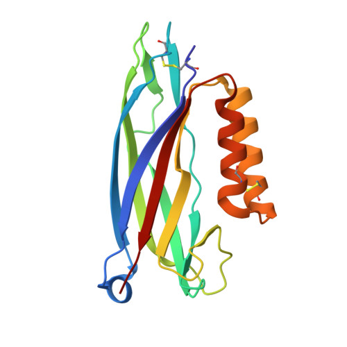
The Yeh pilus adhesin is equipped with an alpha-helical flap motif, which contributes to pectin adherence.

Tamadonfar, K.O.Klein, R.D.Lopatto, E.D.B.Zimmerman, M.I.Azimzadeh, P.N.Sanick, D.A.Porter, J.R.Villicana, J.B.Pinkner, J.S.Chiang, B.H.Gualberto, N.C.Dodson, K.W.Patnode, M.L.Birchenough, G.M.H.Bowman, G.R.Hultgren, S.J.

(2026) Sci Adv 12: eadz1301-eadz1301

  • DOI: https://doi.org/10.1126/sciadv.adz1301
  • Primary Citation of Related Structures:  
    8V9V, 9N4G, 9N4H, 9N4I

  • PubMed Abstract: 

    The Yeh chaperone-usher pathway (CUP) pilus adhesin is encoded in one-half of all Escherichia coli . Yet little is known about its structure and function in E. coli persistence and pathogenesis. Structural investigations reveal that the adhesin receptor binding domains (RBDs) of YehD and its relative YhlD both share a canonical β-rich core and an α-helical flap motif that is hinged at the distal end of the core. This flap was observed in both open and closed conformations using molecular dynamics simulations. The closed conformation is dependent on a hydrophobic patch of amino acids on the distal end of the flap. Functionally, YehD RBD is able to bind pectin, a polysaccharide ubiquitous in plant material. Mutations that interrupt the closed conformation increase the affinity of the protein to pectin, suggesting that the flap contributes mechanistically to pectin binding. Furthermore, in vivo, the pilus contributes to gastrointestinal (GI) tract colonization in the absence of the type 1 pilus. Hence, we report the ability of YehD to bind pectin representing a possible colonization mechanism of the GI tract via a structurally distinct CUP adhesin.


  • Organizational Affiliation
    • Department of Molecular Microbiology and Center for Women's Infectious Disease Research, Washington University School of Medicine, St. Louis, MO, USA.

Macromolecules
Find similar proteins by:  (by identity cutoff)  |  3D Structure
Entity ID: 1
MoleculeChains Sequence LengthOrganismDetailsImage
YehD protein
A, B
171Escherichia coliMutation(s): 1 
UniProt
Find proteins for P33340 (Escherichia coli (strain K12))
Explore P33340 
Go to UniProtKB:  P33340
Entity Groups  
Sequence Clusters30% Identity50% Identity70% Identity90% Identity95% Identity100% Identity
UniProt GroupP33340
Sequence Annotations
Expand
  • Reference Sequence
Experimental Data & Validation

Experimental Data

  • Method: X-RAY DIFFRACTION
  • Resolution: 1.65 Å
  • R-Value Free:  0.238 (Depositor), 0.235 (DCC) 
  • R-Value Work:  0.202 (Depositor), 0.205 (DCC) 
  • R-Value Observed: 0.204 (Depositor) 
Space Group: P 1 21 1
Unit Cell:
Length ( Å )Angle ( ˚ )
a = 34.852α = 90
b = 92.296β = 91.69
c = 44.939γ = 90
Software Package:
Software NamePurpose
PHENIXrefinement
XDSdata reduction
XDSdata scaling
PHENIXphasing

Structure Validation

View Full Validation Report


Entry History & Funding Information

Deposition Data


Funding OrganizationLocationGrant Number
National Institutes of Health/National Institute Of Allergy and Infectious Diseases (NIH/NIAID)United StatesR37AI048689
National Institutes of Health/National Institute Of Allergy and Infectious Diseases (NIH/NIAID)United StatesR01AI029549
National Institutes of Health/National Institute Of Allergy and Infectious Diseases (NIH/NIAID)United StatesT32 AI007172-40

Revision History  (Full details and data files)

  • Version 1.0: 2025-11-26
    Type: Initial release
  • Version 1.1: 2026-01-21
    Changes: Database references