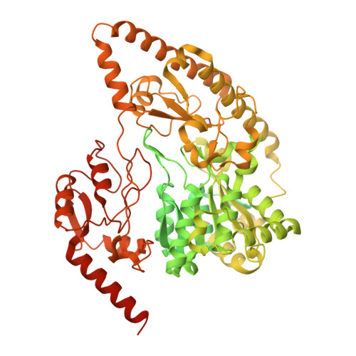
CryoEM structure of Rv2531c reveals cofactor-induced tetramer-dimer transition in a tuberculin amino acid decarboxylase.
Gupta, J., Izard, T.(2025) J Biological Chem 301: 110394
- PubMed: 40543586 Search on PubMedSearch on PubMed Central
- DOI: https://doi.org/10.1016/j.jbc.2025.110394
- Primary Citation Related Structures:
9N0N, 9N0O, 9N0P - PubMed Abstract:
The survival of Mycobacteriumtuberculosis relies on its ability to adapt to dynamic and hostile host environments. Amino acid decarboxylases play a crucial role in these adaptations, but their structural and mechanistic properties are not fully understood. Bioinformatic analyses revealed that these enzymes exist in three distinct forms based on their domain organization. We used cryoEM at 2.76 Å resolution to show that Rv2531c exhibits unexpected oligomeric and conformational flexibility. The enzyme forms a tetramer with distinct open and closed conformations in its apo state, suggesting dynamic intersubunit interactions. Upon binding pyridoxal 5'-phosphate, the enzyme undergoes a dramatic structural rearrangement, transitioning into a dimer. These findings reveal a novel mechanism of oligomeric plasticity. We also uncover an amino-terminal domain that might play a role in this process. Our results provide critical insights into the structural adaptations that support bacterial persistence under intracellular stress. By elucidating the apo and pyridoxal 5'-phosphate-bound states of Rv2531c, we contribute to a deeper understanding of how M. tuberculosis navigates its challenging intracellular environment. These insights into the unique structural features of Rv2531c offer a foundation for targeting metabolic resilience in tuberculosis and open avenues for future studies on the role of this domain in pathogenesis.
- Cell Adhesion Laboratory, UF Scripps, Jupiter, Florida, USA.
Organizational Affiliation: