


Structural and Biophysical Characterization of Thiolato-Cobalamin
Ruetz, M., Mascarenhas, R., Banerjee, R.To be published.
Experimental Data Snapshot
Entity ID: 1 | |||||
|---|---|---|---|---|---|
| Molecule | Chains | Sequence Length | Organism | Details | Image |
| Cyanocobalamin reductase / alkylcobalamin dealkylase | A, C [auth B], D [auth E], F [auth G] | 223 | Homo sapiens | Mutation(s): 0 Gene Names: MMACHC EC: 2.5.1.151 (PDB Primary Data), 1.16.1.6 (PDB Primary Data) | 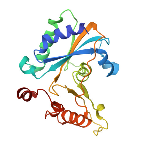 |
UniProt & NIH Common Fund Data Resources | |||||
PHAROS: Q9Y4U1 GTEx: ENSG00000132763 | |||||
Entity Groups | |||||
| Sequence Clusters | 30% Identity50% Identity70% Identity90% Identity95% Identity100% Identity | ||||
| UniProt Group | Q9Y4U1 | ||||
Sequence AnnotationsExpand | |||||
Reference Sequence | |||||
Entity ID: 2 | |||||
|---|---|---|---|---|---|
| Molecule | Chains | Sequence Length | Organism | Details | Image |
| Cobalamin trafficking protein CblD | B [auth D], E [auth F], G [auth C], H | 296 | Homo sapiens | Mutation(s): 0 Gene Names: MMADHC, C2orf25, CL25022, HSPC161, My011 | 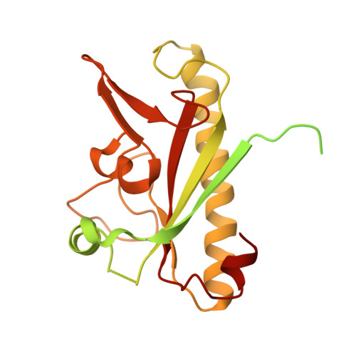 |
UniProt & NIH Common Fund Data Resources | |||||
PHAROS: Q9H3L0 GTEx: ENSG00000168288 | |||||
Entity Groups | |||||
| Sequence Clusters | 30% Identity50% Identity70% Identity90% Identity95% Identity100% Identity | ||||
| UniProt Group | Q9H3L0 | ||||
Sequence AnnotationsExpand | |||||
Reference Sequence | |||||
| Ligands 1 Unique | |||||
|---|---|---|---|---|---|
| ID | Chains | Name / Formula / InChI Key | 2D Diagram | 3D Interactions | |
| B12 (Subject of Investigation/LOI) Download:Ideal Coordinates CCD File | I [auth A], J [auth B], K [auth E], L [auth G] | COBALAMIN C62 H89 Co N13 O14 P LKVIQTCSMMVGFU-DWSMJLPVSA-N |  | ||
| Length ( Å ) | Angle ( ˚ ) |
|---|---|
| a = 72.764 | α = 90 |
| b = 67.983 | β = 99.38 |
| c = 192.66 | γ = 90 |
| Software Name | Purpose |
|---|---|
| PHENIX | refinement |
| Aimless | data scaling |
| XDS | data reduction |
| PHASER | phasing |
| Funding Organization | Location | Grant Number |
|---|---|---|
| National Institutes of Health/National Institute of General Medical Sciences (NIH/NIGMS) | United States | DK045776 |


