Structure-guided design of calcium-dependent protein kinase 1 (CDPK1) inhibitors for cryptosporidiosis.
Wijitrmektong, W., Choi, R., Hulverson, M.A., Liu, L., Cooper, A., Battaile, K.P., Harmon, E.K., Dayao, D.A., Huerta, L., Tzipori, S., McNamara, C.W., Bakowski, M.A., Lovell, S., Van Voorhis, W.C., Cuny, G.D.(2026) J Infect Dis
- PubMed: 42081373 Search on PubMed
- DOI: https://doi.org/10.1093/infdis/jiag242
- Primary Citation Related Structures:
9D8S, 9MI4, 9NTR, 9NTX - PubMed Abstract:
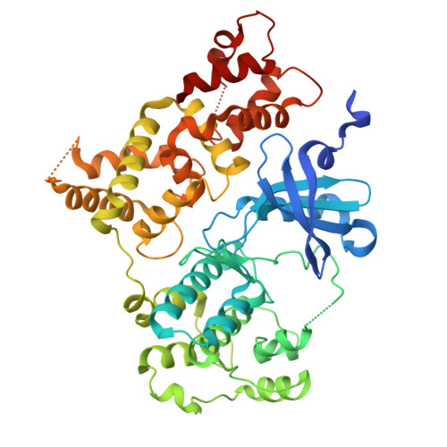
Calcium-dependent protein kinase 1 (CDPK1) has emerged as a protozoan specific target for the treatment of cryptosporidiosis. A previous study identified pyridopyrimidinones as new Cryptosporidium parvum (Cp) CDPK1 inhibitors with potent growth inhibition against C. parvum and C. hominis. Docking analyses suggested the unique positioning of the kinase's αC-helix could present refinement opportunities. Compounds designed to optimize the pyridopyrimidinones focused on the back-pocket region predicted to be proximal to the αC-helix, the solvent exposed region and the ATP ribose-binding site. Designed derivatives were synthesized and assessed for CpCDPK1 and Src kinase inhibition and for Cryptosporidium spp., growth inhibition in mammalian cells. AMP/Mg+2 and three inhibitors were co-crystalized with CpCDPK1, and two inhibitors were profiled for kinase selectivity. WIN 4-88 was identified with CpCDPK1 (IC50 = 0.056 μM), and growth inhibition of zoonotic C. parvum (NLuc EC50 = 0.042 μM), anthroponotic C. parvum (Tu114 EC50 = 0.030 μM), and C. hominis Tu502 (EC50 = 0.062 μM), as well as enhanced kinome selectivity. The crystal structures confirmed the predicted binding mode, indicating key interactions with hinge residue Y155, similar orientations of the solvent expose moieties, occupancy of the back-pocket near the αC-helix and for one inhibitor containing a solubilizing hydroxyethyl attached to the central heterocycle extension into the ATP ribose-binding site. The expanded structure-activity relationship and structural insights will potentially be applicable to other chemotypes with similar binding modes and will enhance development of CpCDPK1 inhibitors for the treatment of cryptosporidiosis.
- Department of Pharmacological and Pharmaceutical Sciences, College of Pharmacy, University of Houston, Houston, Texas 77204, United States.
Organizational Affiliation: