Interplay between C alpha Methylation and C alpha Stereochemistry in the Folding Energetics of a Helix-Rich Miniprotein.
Harmon, T.W., Lin, Y., Sutton, R.T., Osborne, S.W.J., Seth Horne, W.(2025) Chembiochem 26: e202401022-e202401022
- PubMed: 39791987 Search on PubMed
- DOI: https://doi.org/10.1002/cbic.202401022
- Primary Citation Related Structures:
9MF7, 9MF8, 9MF9, 9MFA, 9MFB - PubMed Abstract:
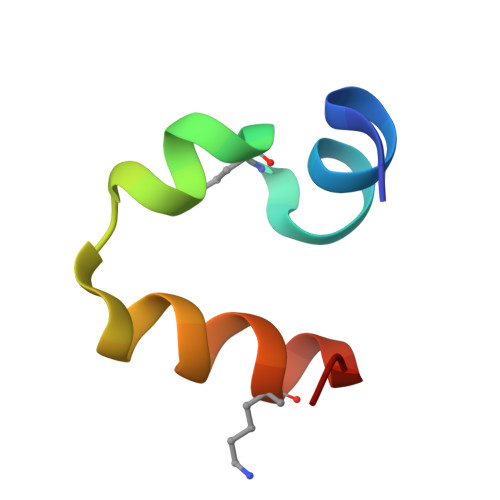
The α-helix is an abundant and functionally important element of protein secondary structure, which has motivated intensive efforts toward chemical strategies to stabilize helical folds. One such method is the incorporation of non-canonical backbone composition through an additional methyl substituent at the C α atom. Examples of monomers include the achiral 2-aminoisobutyric acid (Aib) with geminal dimethyl substitution and chiral analogues with one methyl and one non-methyl substituent. While Aib and chiral C α -Me residues are both established helix promoting moieties, their comparative ability in this regard has not been quantitatively investigated. Addressing this gap would help to inform the use of these building blocks in the construction of peptide and protein mimetics as well as provide fundamental insights into consequences of backbone methylation on folding. Here, we report a quantitative comparison of the impacts of Aib and chiral αMe residues on the high-resolution folded structure and folding thermodynamics of a small helical protein. These results reveal a synergistic stabilizing effect arising from the presence of C α methylation in conjunction with a C α stereocenter.
- Department of Chemistry, University of Pittsburgh, Pittsburgh, PA, 15260, USA.
Organizational Affiliation: