


SIDT1 is a calcium-associated CD8+ T cell suppressor and target for cancer immunotherapy
Hu, T., Han, Z., Liao, L., Zeng, Y., Ma, H., Huang, J., Chen, Y., Ye, J., Zhou, T., Zhang, W., Cang, C., Sun, L., Pan, W., Wang, J., Zhu, S.To be published.
Experimental Data Snapshot
wwPDB Validation 3D Report Full Report

Entity ID: 1 | |||||
|---|---|---|---|---|---|
| Molecule | Chains | Sequence Length | Organism | Details | Image |
| Isoform 2 of SID1 transmembrane family member 1 | 832 | Homo sapiens | Mutation(s): 0 Gene Names: SIDT1 | 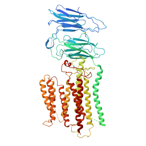 | |
UniProt & NIH Common Fund Data Resources | |||||
GTEx: ENSG00000072858 | |||||
Entity Groups | |||||
| Sequence Clusters | 30% Identity50% Identity70% Identity90% Identity95% Identity100% Identity | ||||
| UniProt Group | Q9NXL6 | ||||
Sequence AnnotationsExpand | |||||
Reference Sequence | |||||
| Task | Software Package | Version |
|---|---|---|
| MODEL REFINEMENT | PHENIX | 1.13_2998: |
| Funding Organization | Location | Grant Number |
|---|---|---|
| Chinese Academy of Sciences | China | -- |


