9LYP | pdb_00009lyp

Alpha SARS-CoV-2 spike protein RBD-down in complex with REGN10987 Fab homologue (local refinement)


Experimental Data Snapshot

  • Method: ELECTRON MICROSCOPY
  • Resolution: 3.60 Å
  • Aggregation State: PARTICLE 
  • Reconstruction Method: SINGLE PARTICLE 

wwPDB Validation   3D Report Full Report


This is version 1.0 of the entry. See complete history


Literature

Structure and dynamics of Alpha B.1.1.7 SARS-CoV-2 S-protein in complex with Fab of neutralizing antibody REGN10987.

Kocharovskaya, M.V.Pichkur, E.B.Ivannikov, A.D.Kharlampieva, D.D.Grafskaia, E.N.Lyukmanova, E.N.Kirpichnikov, M.P.Shenkarev, Z.O.

(2025) Biochem Biophys Res Commun 755: 151558-151558

  • DOI: https://doi.org/10.1016/j.bbrc.2025.151558
  • Primary Citation of Related Structures:  
    9LYO, 9LYP

  • PubMed Abstract: 

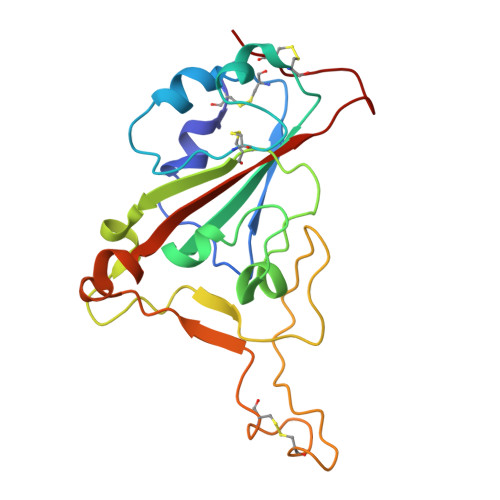
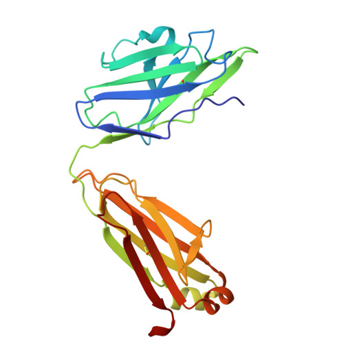
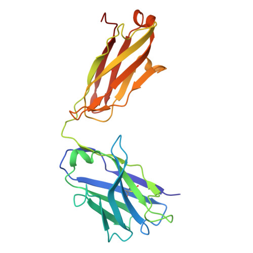
    One of the approaches for treatment of COVID-19 is a use of neutralizing antibodies (nAbs). The study of the mechanisms by which nAbs recognize different strains of SARS-CoV-2 may facilitate the development of new drugs and vaccines against the coronavirus infection. In this work, we present the 3.1 Å resolution cryo-electron microscopy structure of a full-length trimeric spike-protein (S-protein) of the SARS-CoV-2 Alpha (B.1.1.7) variant in complex with the Fab of the REGN10987 nAb. In the complex, two receptor-binding domains (RBDs) of the S-protein were observed in the 'up' state, whereas third RBD was in the 'down' state. This distinguishes the obtained structure from the complex of Delta (B.1.617.2) S-protein with REGN10987-Fab, where only one RBD was in the 'up' state. Probably some of the substituted residues (K478T, A570D, and S982A) located at the interprotomer interfaces are responsible for the greater Alpha S-protein opening upon the REGN10987-Fab binding. The Fab identically binds to the RBD in the both 'up' and 'down' conformations. The RBD-Fab interaction interface was refined to a resolution of 3.6 Å. The antibody binds to the receptor-binding motif (RBM), which prevents the S-protein from the binding to its receptor, angiotensin-converting enzyme 2 (ACE-2). Comparison with the structures of the Wuhan (wild type) and Delta RBD variants in complex with REGN10987-Fab revealed that the N501Y and T478K/L452R mutations presented in the RBM of the Alpha and Delta variants, respectively, do not affect the mode of the RBD-Fab interaction.


  • Organizational Affiliation
    • Department of Structural Biology, Shemyakin-Ovchinnikov Institute of Bioorganic Chemistry, Russian Academy of Sciences, 117997, Moscow, Russia; Moscow Center for Advanced Studies, 123592, Moscow, Russia.

Macromolecules
Find similar proteins by:  (by identity cutoff)  |  3D Structure
Entity ID: 1
MoleculeChains Sequence LengthOrganismDetailsImage
Spike glycoproteinA [auth B]194Severe acute respiratory syndrome coronavirus 2Mutation(s): 0 
UniProt
Find proteins for A0A6V7ALX9 (Severe acute respiratory syndrome coronavirus 2)
Explore A0A6V7ALX9 
Go to UniProtKB:  A0A6V7ALX9
Entity Groups  
Sequence Clusters30% Identity50% Identity70% Identity90% Identity95% Identity100% Identity
UniProt GroupA0A6V7ALX9
Glycosylation
Glycosylation Sites: 1
Sequence Annotations
Expand
  • Reference Sequence
Find similar proteins by:  (by identity cutoff)  |  3D Structure
Entity ID: 2
MoleculeChains Sequence LengthOrganismDetailsImage
REGN10987 Fab homologue (Light chain)B [auth i]218Homo sapiensMutation(s): 0 
Entity Groups  
Sequence Clusters30% Identity50% Identity70% Identity90% Identity95% Identity100% Identity
Sequence Annotations
Expand
  • Reference Sequence
Find similar proteins by:  (by identity cutoff)  |  3D Structure
Entity ID: 3
MoleculeChains Sequence LengthOrganismDetailsImage
REGN10987 Fab homologue (Heavy chain)C [auth j]223Homo sapiensMutation(s): 0 
Entity Groups  
Sequence Clusters30% Identity50% Identity70% Identity90% Identity95% Identity100% Identity
Sequence Annotations
Expand
  • Reference Sequence
Oligosaccharides

Help

Entity ID: 4
MoleculeChains Length2D Diagram Glycosylation3D Interactions
2-acetamido-2-deoxy-beta-D-glucopyranose-(1-4)-2-acetamido-2-deoxy-beta-D-glucopyranoseD [auth P]2N-Glycosylation
Glycosylation Resources
GlyTouCan:  G42666HT
GlyCosmos:  G42666HT
GlyGen:  G42666HT
Experimental Data & Validation

Experimental Data

  • Method: ELECTRON MICROSCOPY
  • Resolution: 3.60 Å
  • Aggregation State: PARTICLE 
  • Reconstruction Method: SINGLE PARTICLE 
EM Software:
TaskSoftware PackageVersion
MODEL REFINEMENTPHENIX1.20.1_4487:

Structure Validation

View Full Validation Report


Entry History & Funding Information

Deposition Data


Funding OrganizationLocationGrant Number
Not funded--

Revision History  (Full details and data files)

  • Version 1.0: 2025-04-02
    Type: Initial release