9LUY | pdb_00009luy

Single-chain Fv antibody of G2 fused with antigen peptide from chicken chromosome 6 C10orf76 homolog


Experimental Data Snapshot

  • Method: X-RAY DIFFRACTION
  • Resolution: 1.62 Å
  • R-Value Free: 
    0.188 (Depositor), 0.188 (DCC) 
  • R-Value Work: 
    0.167 (Depositor), 0.167 (DCC) 
  • R-Value Observed: 
    0.168 (Depositor) 

Starting Model: experimental
View more details

wwPDB Validation 3D Report Full Report

Validation slider image for 9LUY

This is version 1.1 of the entry. See complete history

Literature

Crystal structures reveal how the multispecific antibody G2 achieves binding to different peptides.

Hanazono, Y.Yabuno, S.Hayashi, T.Numoto, N.Kamatari, Y.O.Ito, N.Oda, M.

(2025) FEBS Lett 

  • DOI: https://doi.org/10.1002/1873-3468.70095
  • Primary Citation Related Structures: 
    9LUX, 9LUY

  • PubMed Abstract: 

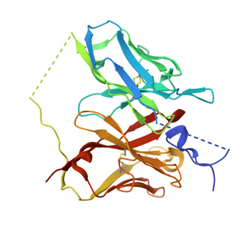
    Monoclonal antibody G2, obtained via immunization with the chicken prion protein (ChPrP), has unique antigen-binding specificity. G2 specifically binds to the ChPrP-derived peptide (Pep18mer) as expected, but also to three other peptides. In this study, we determined the crystal structures of G2 single-chain Fv antibodies covalently linked to Pep18mer and another peptide, PepH4P6. Both bound peptides formed similar U-shaped structures that stuck into the G2 antigen-binding pocket. Their three-dimensional structures were stabilized by interactions within the peptides, and the structure of the bound Pep18mer was similar to that of the corresponding ChPrP region. G2 acquired the binding ability to both Pep18mer and PepH4P6 via deletion of the 95th residue of the light chain during affinity maturation, consistent with our structural analysis.


  • Organizational Affiliation
    • Medical Research Laboratory, Institute of Integrated Research, Institute of Science Tokyo, Japan.

Macromolecule Content 

  • Total Structure Weight: 29.21 kDa 
  • Atom Count: 2,100 
  • Modeled Residue Count: 242 
  • Deposited Residue Count: 273 
  • Unique protein chains: 1

Macromolecules

Find similar proteins by:|  3D Structure
Entity ID: 1
MoleculeChains  Sequence LengthOrganismDetailsImage
Single-chain Fv antibody of G2 fused with antigen peptide from chicken chromosome 6 C10orf76 homolog273Gallus gallusMus musculus
This entity is chimeric
Mutation(s): 0 

Small Molecules

Ligands 1 Unique
IDChains Name / Formula / InChI Key2D Diagram3D Interactions
GOL

Query on GOL


Download:Ideal Coordinates CCD File
B [auth A]GLYCEROL
C3 H8 O3
PEDCQBHIVMGVHV-UHFFFAOYSA-N

Experimental Data & Validation

Experimental Data

  • Method: X-RAY DIFFRACTION
  • Resolution: 1.62 Å
  • R-Value Free:  0.188 (Depositor), 0.188 (DCC) 
  • R-Value Work:  0.167 (Depositor), 0.167 (DCC) 
  • R-Value Observed: 0.168 (Depositor) 
Space Group: C 1 2 1
Unit Cell:
Length ( Å )Angle ( ˚ )
a = 76.054α = 90
b = 71.39β = 100.908
c = 43.352γ = 90
Software Package:
Software NamePurpose
PHENIXrefinement
XDSdata reduction
XDSdata scaling
PHASERphasing

Structure Validation

View Full Validation Report


Entry History 

& Funding Information

Deposition Data


Funding OrganizationLocationGrant Number
Not funded--

Revision History  (Full details and data files)

  • Version 1.0: 2025-06-11
    Type: Initial release
  • Version 1.1: 2025-07-02
    Changes: Database references