9LSZ | pdb_00009lsz

Inward-open Structure of human B0AT1


Experimental Data Snapshot

  • Method: ELECTRON MICROSCOPY
  • Resolution: 3.11 Å
  • Aggregation State: PARTICLE 
  • Reconstruction Method: SINGLE PARTICLE 

wwPDB Validation 3D Report Full Report

Validation slider image for 9LSZ

This is version 1.0 of the entry. See complete history

Literature

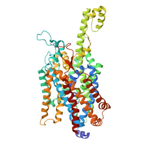
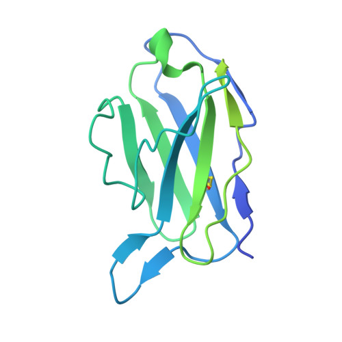
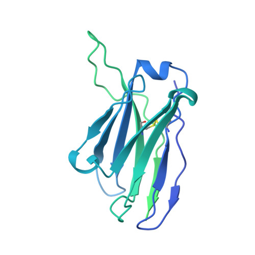
Structure-guided development of a potent human B0AT1 inhibitor effective in a mouse model of phenylketonuria

Imazu, T.Akashi, T.Hiraizumi, M.Inui, Y.Sasaki, W.Takahashi, T.Todoroki, H.Kumanomidou, T.Hisano, H.Asada, H.Kusakizako, T.Nishizawa, T.Iwata, S.Nureki, O.Miyaguchi, I.

To be published.

Macromolecule Content 

  • Total Structure Weight: 130.67 kDa 
  • Atom Count: 6,458 
  • Modeled Residue Count: 813 
  • Deposited Residue Count: 1,174 
  • Unique protein chains: 3

Macromolecules

Find similar proteins by:|  3D Structure
Entity ID: 1
MoleculeChains  Sequence LengthOrganismDetailsImage
Sodium-dependent neutral amino acid transporter B(0)AT1614Homo sapiensMutation(s): 0 
Gene Names: SLC6A19B0AT1
UniProt & NIH Common Fund Data Resources
Find proteins for Q695T7 (Homo sapiens)
Explore Q695T7 
Go to UniProtKB:  Q695T7
PHAROS:  Q695T7
GTEx:  ENSG00000174358 
Entity Groups
Sequence Clusters30% Identity50% Identity70% Identity90% Identity95% Identity100% Identity
UniProt GroupQ695T7
Glycosylation
Glycosylation Sites: 1Go to GlyGen: Q695T7-1
Sequence Annotations
Expand
Reference Sequence
Find similar proteins by:|  3D Structure
Entity ID: 2
MoleculeChains  Sequence LengthOrganismDetailsImage
Antibody Fab Light chainB [auth L]225Mus musculusMutation(s): 0 
Find similar proteins by:|  3D Structure
Entity ID: 3
MoleculeChains  Sequence LengthOrganismDetailsImage
Antibody Fab Heavy chainC [auth H]335Mus musculusMutation(s): 0 

Oligosaccharides

Help  
Entity ID: 4
MoleculeChains Length2D Diagram GlycosylationD Interactions
2-acetamido-2-deoxy-beta-D-glucopyranose-(1-4)-2-acetamido-2-deoxy-beta-D-glucopyranoseD [auth B]2N-Glycosylation
Glycosylation Resources
GlyTouCan: G42666HT
GlyCosmos: G42666HT
GlyGen: G42666HT

Experimental Data & Validation

Experimental Data

  • Method: ELECTRON MICROSCOPY
  • Resolution: 3.11 Å
  • Aggregation State: PARTICLE 
  • Reconstruction Method: SINGLE PARTICLE 
EM Software:
TaskSoftware PackageVersion
MODEL REFINEMENTREFMAC5.8.0267

Structure Validation

View Full Validation Report


Entry History 

& Funding Information

Deposition Data


Funding OrganizationLocationGrant Number
Not funded--

Revision History  (Full details and data files)

  • Version 1.0: 2026-02-18
    Type: Initial release