9LKP | pdb_00009lkp

X-ray structure of Enterobacter cloaca transaldolase


Experimental Data Snapshot

  • Method: X-RAY DIFFRACTION
  • Resolution: 1.63 Å
  • R-Value Free: 
    0.197 (Depositor), 0.204 (DCC) 
  • R-Value Work: 
    0.173 (Depositor), 0.185 (DCC) 
  • R-Value Observed: 
    0.175 (Depositor) 

Starting Model: experimental
View more details

wwPDB Validation 3D Report Full Report

Validation slider image for 9LKP

This is version 1.1 of the entry. See complete history

Literature

Synthetic Study of 8- and 9-Carbon Sugars by Transaldolase.

Yoshihara, A.Miyoshi, E.Tomino, S.Hanaki, Y.Mochizuki, S.Yoshida, H.Izumori, K.Kamitori, S.

(2025) J Agric Food Chem 73: 18914-18922

  • DOI: https://doi.org/10.1021/acs.jafc.5c05539
  • Primary Citation Related Structures: 
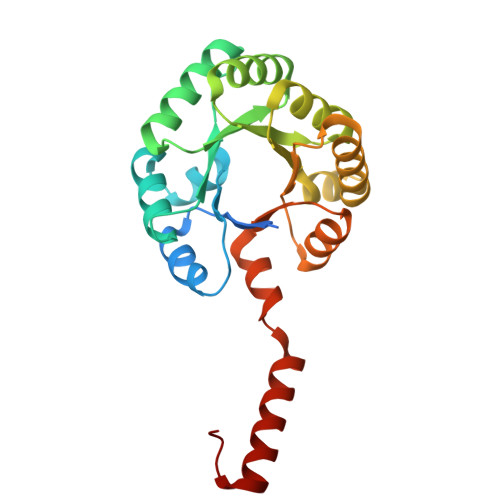
    9LKP, 9LL3, 9UI2

  • PubMed Abstract: 

    In nature, higher carbon sugars composed of 7 or more carbons exist in limited quantities. Since some higher carbon sugars have attracted attention due to their biological activities, it is necessary to develop a strategy to synthesize them. Transaldolase catalyzes the transfer of three-carbon units from d-fructose-6-phosphate (donor) to d-erythrulose-4-phosphate (acceptor) to produce d-sedoheptulose-7-phosphate. If transaldolase can recognize nonphosphorylated monosaccharides as substrates, it can synthesize 8-carbon octuloses and 9-carbon nonuloses using nonphosphorylated pentoses and hexoses as acceptors, respectively. We performed biochemical and structural characterization of thermophilic Thermus thermophilus HB8 transaldolase and successfully synthesized octuloses and nonuloses using nonphosphorylated aldoses as acceptors: d-ribose (conversion rate of 74%), d-xylose (55%), l-arabinose (49%), l-lyxose (84%), d-allose (13%), d-galactose (56%), and l-altrose (71%). Products were identified by LC/MS and NMR spectroscopic analyses. X-ray structure of the enzyme showed that the wide and hydrophilic catalytic site facilitates the binding of nonphosphorylated aldoses as acceptors.


  • Organizational Affiliation
    • International Institute of Rare Sugar Research and Education, Kagawa University, Takamatsu, Kagawa 760-8521, Japan.

Macromolecule Content 

  • Total Structure Weight: 126.66 kDa 
  • Atom Count: 8,925 
  • Modeled Residue Count: 1,100 
  • Deposited Residue Count: 1,205 
  • Unique protein chains: 1

Macromolecules

Find similar proteins by:|  3D Structure
Entity ID: 1
MoleculeChains  Sequence LengthOrganismDetailsImage
Fructose-6-phosphate aldolase
A, B, C, D, E
241Enterobacter cloacaeMutation(s): 0 
Gene Names: fsaDP202_18755
EC: 4.1.2
UniProt
Find proteins for A0A6G4MPG4 (Enterobacter hormaechei)
Explore A0A6G4MPG4 
Go to UniProtKB:  A0A6G4MPG4
Entity Groups
Sequence Clusters30% Identity50% Identity70% Identity90% Identity95% Identity100% Identity
UniProt GroupA0A6G4MPG4
Sequence Annotations
Expand
Reference Sequence

Experimental Data & Validation

Experimental Data

  • Method: X-RAY DIFFRACTION
  • Resolution: 1.63 Å
  • R-Value Free:  0.197 (Depositor), 0.204 (DCC) 
  • R-Value Work:  0.173 (Depositor), 0.185 (DCC) 
  • R-Value Observed: 0.175 (Depositor) 
Space Group: C 2 2 21
Unit Cell:
Length ( Å )Angle ( ˚ )
a = 99α = 90
b = 180.07β = 90
c = 134.04γ = 90
Software Package:
Software NamePurpose
REFMACrefinement
XDSdata reduction
XDSdata scaling
MOLREPphasing

Structure Validation

View Full Validation Report


Entry History 

& Funding Information

Deposition Data

  • Released Date: 2025-07-30 
  • Deposition Author(s): Kamitori, S.

Funding OrganizationLocationGrant Number
Not funded--

Revision History  (Full details and data files)

  • Version 1.0: 2025-07-30
    Type: Initial release
  • Version 1.1: 2025-08-06
    Changes: Database references