9LIP | pdb_00009lip

The cryo-EM structure of the native PMEL fibril lamella


Experimental Data Snapshot

  • Method: ELECTRON MICROSCOPY
  • Resolution: 3.48 Å
  • Aggregation State: FILAMENT 
  • Reconstruction Method: SINGLE PARTICLE 

wwPDB Validation   3D Report Full Report


This is version 1.0 of the entry. See complete history


Literature

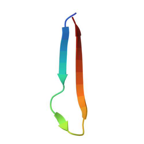
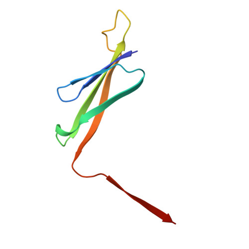
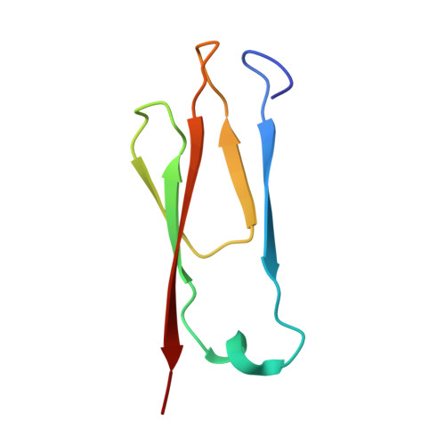
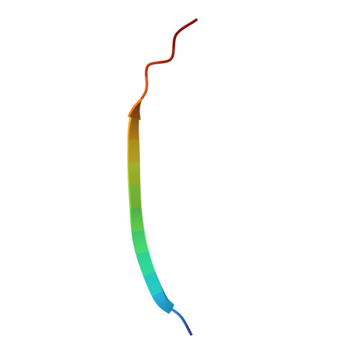
Atomic structure and in situ visualization of native PMEL lamellae in melanosomes.

Ma, B.Yao, Y.Dong, H.Yang, L.Li, D.Zhao, Q.Sun, B.Chen, Y.Liu, C.Li, D.

(2025) Nat Commun 16: 10300-10300

  • DOI: https://doi.org/10.1038/s41467-025-65221-0
  • Primary Citation of Related Structures:  
    9LIP

  • PubMed Abstract: 

    Melanin synthesis within melanosomes critically depends on the fibrillar architecture formed by the pigment cell-specific protein PMEL. Although PMEL fibrils have historically been classified as functional amyloids, their native supramolecular organization in situ and detailed molecular architecture have remained unresolved. In this study, we combine in situ cryo-electron tomography (cryo-ET) and cryo-electron microscopy (cryo-EM) to elucidate the native structural organization of PMEL fibrils within human melanosomes from both patient melanoma tissues and melanocyte cell lines. We demonstrate that PMEL does not form conventional isolated amyloid fibrils, but rather assembles into highly organized lamellar sheets consisting of laterally aligned fibrils interconnected by flexible linker regions. Cryo-EM structures reveal a distinctive butterfly-shaped fibril unit composed of multiple structured domains, including both the proteolytically cleaved Mα and Mβ fragments of PMEL, which assemble into a amyloid-like β-sheet arrangement. Notably, we identify intrinsically disordered regions critical for lamellar assembly and curvature and verify key glycosylation modifications in the structure. This architecture distinguishes PMEL fibrils from canonical amyloids and elucidates the molecular basis underlying melanosome integrity and pigmentation. Moreover, our work provides molecular insights relevant for pigmentation disorders and PMEL-associated diseases, including melanoma.


  • Organizational Affiliation
    • Interdisciplinary Research Center on Biology and Chemistry, State Key Laboratory of Chemical Biology, Shanghai Institute of Organic Chemistry, Chinese Academy of Sciences, Shanghai, China.

Macromolecules

Find similar proteins by:  Sequence   |   3D Structure  

Entity ID: 1
MoleculeChains Sequence LengthOrganismDetailsImage
M-alpha24Homo sapiensMutation(s): 0 
UniProt & NIH Common Fund Data Resources
Find proteins for P40967 (Homo sapiens)
Explore P40967 
Go to UniProtKB:  P40967
PHAROS:  P40967
GTEx:  ENSG00000185664 
Entity Groups  
Sequence Clusters30% Identity50% Identity70% Identity90% Identity95% Identity100% Identity
UniProt GroupP40967
Sequence Annotations
Expand
  • Reference Sequence
Find similar proteins by:  (by identity cutoff)  |  3D Structure
Entity ID: 2
MoleculeChains Sequence LengthOrganismDetailsImage
M-alpha75Homo sapiensMutation(s): 0 
UniProt & NIH Common Fund Data Resources
Find proteins for P40967 (Homo sapiens)
Explore P40967 
Go to UniProtKB:  P40967
PHAROS:  P40967
GTEx:  ENSG00000185664 
Entity Groups  
Sequence Clusters30% Identity50% Identity70% Identity90% Identity95% Identity100% Identity
UniProt GroupP40967
Sequence Annotations
Expand
  • Reference Sequence
Find similar proteins by:  (by identity cutoff)  |  3D Structure
Entity ID: 3
MoleculeChains Sequence LengthOrganismDetailsImage
M-alpha68Homo sapiensMutation(s): 0 
UniProt & NIH Common Fund Data Resources
Find proteins for P40967 (Homo sapiens)
Explore P40967 
Go to UniProtKB:  P40967
PHAROS:  P40967
GTEx:  ENSG00000185664 
Entity Groups  
Sequence Clusters30% Identity50% Identity70% Identity90% Identity95% Identity100% Identity
UniProt GroupP40967
Sequence Annotations
Expand
  • Reference Sequence

Find similar proteins by:  Sequence   |   3D Structure  

Entity ID: 4
MoleculeChains Sequence LengthOrganismDetailsImage
M-beta17Homo sapiensMutation(s): 0 
UniProt & NIH Common Fund Data Resources
Find proteins for P40967 (Homo sapiens)
Explore P40967 
Go to UniProtKB:  P40967
PHAROS:  P40967
GTEx:  ENSG00000185664 
Entity Groups  
Sequence Clusters30% Identity50% Identity70% Identity90% Identity95% Identity100% Identity
UniProt GroupP40967
Sequence Annotations
Expand
  • Reference Sequence
Experimental Data & Validation

Experimental Data

  • Method: ELECTRON MICROSCOPY
  • Resolution: 3.48 Å
  • Aggregation State: FILAMENT 
  • Reconstruction Method: SINGLE PARTICLE 
EM Software:
TaskSoftware PackageVersion
MODEL REFINEMENTPHENIX

Structure Validation

View Full Validation Report


Entry History & Funding Information

Deposition Data


Funding OrganizationLocationGrant Number
Not funded--

Revision History  (Full details and data files)

  • Version 1.0: 2025-12-24
    Type: Initial release