9LEW | pdb_00009lew

The crystal structure of DinJ-YafQ complex from Vibrio cholerae


Experimental Data Snapshot

  • Method: X-RAY DIFFRACTION
  • Resolution: 2.30 Å
  • R-Value Free: 
    0.231 (Depositor), 0.229 (DCC) 
  • R-Value Work: 
    0.201 (Depositor), 0.201 (DCC) 
  • R-Value Observed: 
    0.202 (Depositor) 

Starting Model: in silico
View more details

wwPDB Validation 3D Report Full Report

Validation slider image for 9LEW

Ligand Structure Quality Assessment 


This is version 1.0 of the entry. See complete history

Literature

Novel oligomerization state of DinJ-YafQ complex from Vibrio cholerae-Structural and biochemical insights.

Mun, S.A.Yeon, G.B.Park, Y.H.Yoon, H.Ha, J.H.Jang, T.H.Hong, E.Bae, J.Son, J.

(2025) Int J Biol Macromol 320: 145865-145865

  • DOI: https://doi.org/10.1016/j.ijbiomac.2025.145865
  • Primary Citation Related Structures: 
    9LEW

  • PubMed Abstract: 

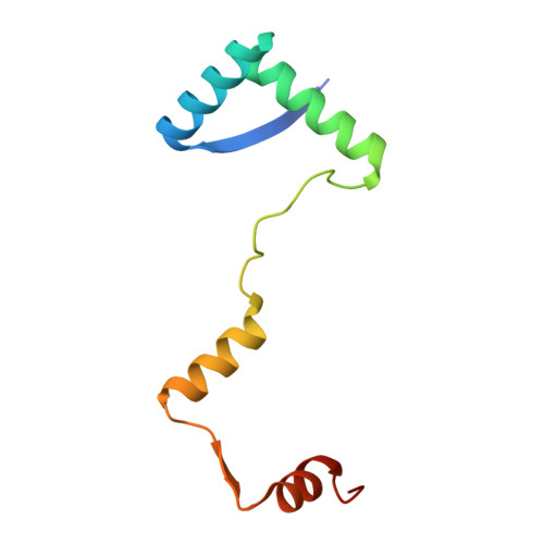
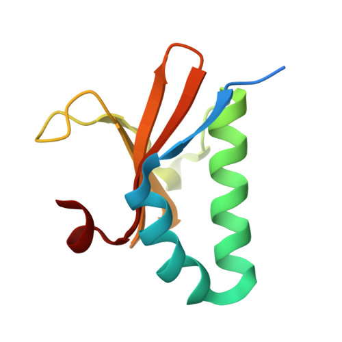
    Toxin-antitoxin (TA) systems are widely distributed in archaeal and bacterial genomes and are crucial for maintaining the physiological functions required for cellular homeostasis. The DinJ-YafQ TA system belongs to the well-known RelBE family of Type II TA systems. Vibrio cholerae, the causative agent of cholera, infects humans through the consumption of contaminated, unpurified drinking water. Its genome contains a cassette of 19 TA system-related genes located on chromosome II, six of which are classified under the RelBE family. Herein, we determined the crystal structure of the V. cholerae DinJ (VcDinJ, antitoxin)-YafQ (VcYafQ, toxin) complex at 2.3 Å resolution. The unique orientation of the VcDinJ RHH 2 motifs relative to the YafQ molecules promotes the formation of a highly oligomerized hetero-hexadecamer (16-mer) in a concentration-dependent manner. According to biochemical experiments, the VcDinJ-YafQ complex directly binds to the promoter region of its operon, suggesting its role as a transcriptional repressor. An autoregulatory mechanism in which DNA binding induces conformational changes is suggested based on structural modeling through DNA superposition. This finding supports the concentration-dependent and DNA-associated oligomerization of the VcDinJ-YafQ complex.


  • Organizational Affiliation
    • New Drug Development Center, Daegu-Gyeongbuk Medical Innovation Foundation (DGMIF), 360-4 Dongnae-dong, Dong-gu, Daegu 41061, Republic of Korea.

Macromolecule Content 

  • Total Structure Weight: 87.68 kDa 
  • Atom Count: 5,862 
  • Modeled Residue Count: 712 
  • Deposited Residue Count: 776 
  • Unique protein chains: 2

Macromolecules

Find similar proteins by:|  3D Structure
Entity ID: 1
MoleculeChains  Sequence LengthOrganismDetailsImage
DNA-damage-inducible protein J
A, C, E, G
94Vibrio cholerae O1 biovar El Tor str. N16961Mutation(s): 0 
Gene Names: VC_A0324
UniProt
Find proteins for Q9KML3 (Vibrio cholerae serotype O1 (strain ATCC 39315 / El Tor Inaba N16961))
Explore Q9KML3 
Go to UniProtKB:  Q9KML3
Entity Groups
Sequence Clusters30% Identity50% Identity70% Identity90% Identity95% Identity100% Identity
UniProt GroupQ9KML3
Sequence Annotations
Expand
Reference Sequence
Find similar proteins by:|  3D Structure
Entity ID: 2
MoleculeChains  Sequence LengthOrganismDetailsImage
Type II toxin-antitoxin system YafQ family toxin
B, D, F, H
100Vibrio cholerae O1 biovar El Tor str. N16961Mutation(s): 1 
Gene Names: VC_A0323
UniProt
Find proteins for Q9KML4 (Vibrio cholerae serotype O1 (strain ATCC 39315 / El Tor Inaba N16961))
Explore Q9KML4 
Go to UniProtKB:  Q9KML4
Entity Groups
Sequence Clusters30% Identity50% Identity70% Identity90% Identity95% Identity100% Identity
UniProt GroupQ9KML4
Sequence Annotations
Expand
Reference Sequence

Small Molecules

Ligands 2 Unique
IDChains Name / Formula / InChI Key2D Diagram3D Interactions
SO4

Query on SO4


Download:Ideal Coordinates CCD File
I [auth B]SULFATE ION
O4 S
QAOWNCQODCNURD-UHFFFAOYSA-L
ACT
(Subject of Investigation/LOI)

Query on ACT


Download:Ideal Coordinates CCD File
J [auth C]
K [auth C]
L [auth D]
M [auth D]
N [auth D]
J [auth C],
K [auth C],
L [auth D],
M [auth D],
N [auth D],
O [auth E],
P [auth H],
Q [auth H]
ACETATE ION
C2 H3 O2
QTBSBXVTEAMEQO-UHFFFAOYSA-M

Experimental Data & Validation

Experimental Data

  • Method: X-RAY DIFFRACTION
  • Resolution: 2.30 Å
  • R-Value Free:  0.231 (Depositor), 0.229 (DCC) 
  • R-Value Work:  0.201 (Depositor), 0.201 (DCC) 
  • R-Value Observed: 0.202 (Depositor) 
Space Group: P 62 2 2
Unit Cell:
Length ( Å )Angle ( ˚ )
a = 163.924α = 90
b = 163.924β = 90
c = 114.035γ = 120
Software Package:
Software NamePurpose
PHENIXrefinement
XDSdata reduction
XSCALEdata scaling
PHASERphasing

Structure Validation

View Full Validation Report


Ligand Structure Quality Assessment 


Entry History 

& Funding Information

Deposition Data


Funding OrganizationLocationGrant Number
National Research Foundation (NRF, Korea)Korea, Republic OfRS-2024-00398456

Revision History  (Full details and data files)

  • Version 1.0: 2025-07-30
    Type: Initial release